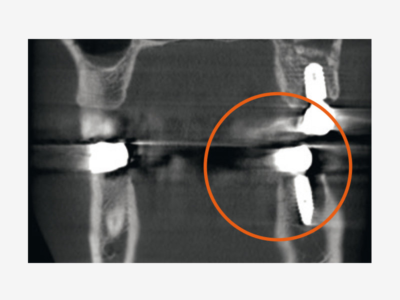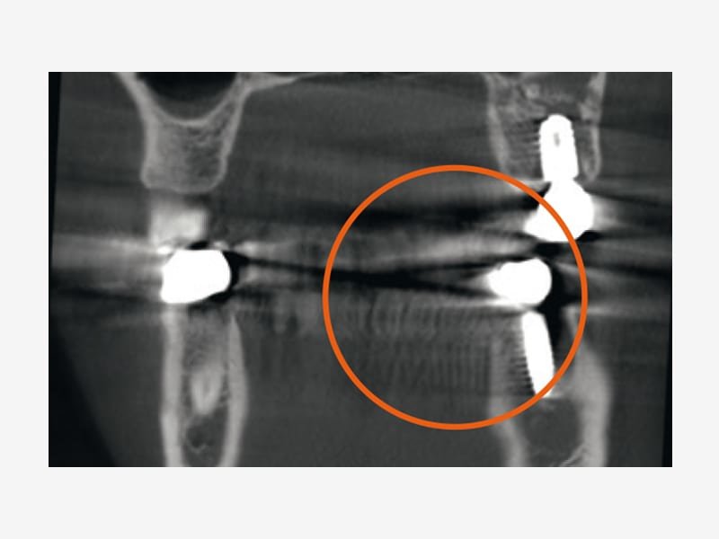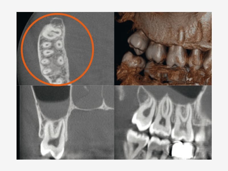
Sharp imaging plays a key role in patient health. The sharper the image, the more accurate the diagnosis – resulting in a more predictable and successful treatment.
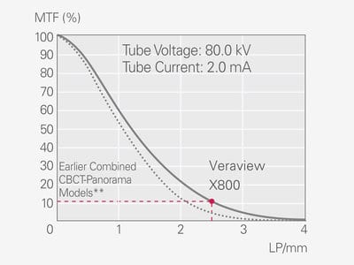
Sharp performance: Peak values of 80 μm and 2.5 LP/mm
This is where the Veraview X800 is especially impressive, with an absolutely top performance and uncompromising resolution. Voxel size for images in the Ø 40 mm × H 40 mm field of view (FOV) is a surprising 80 μm – yielding a brilliant resolution of 2.5 LP/mm. MTF (Modulation Transfer Function) is one way to objectively evaluate the linepair resolution and objectively expresses how many line-pairs and at what level of contrast can be discriminated. Generally, if MTF is 10%, naked eye discrimination is possible. Spatial resolution does not depend only on voxel size.
** Veraviewepocs 3D series
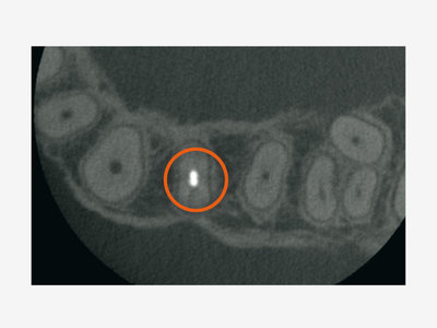
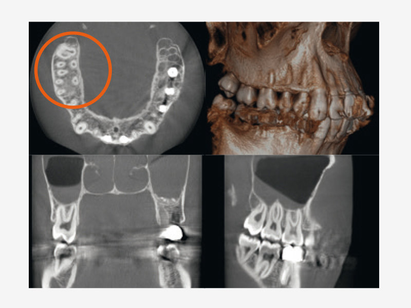
High resolution, voxel size 80 μm
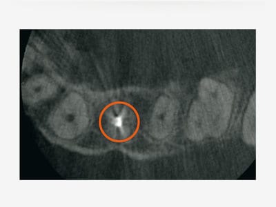
Standard, voxel size 125 μm**