Veraviewepocs 2D

  • Veraviewepocs 2D
  • Technical Data

Panoramic/Cephalometric X-ray

Morita's cutting edge technology features extremely high quality images with low exposure. Veraviewepocs 2D is completely upgradeable to our Veraviewepocs 3D model. This unit also features the NEW High Definition (HD) cephalometric update.

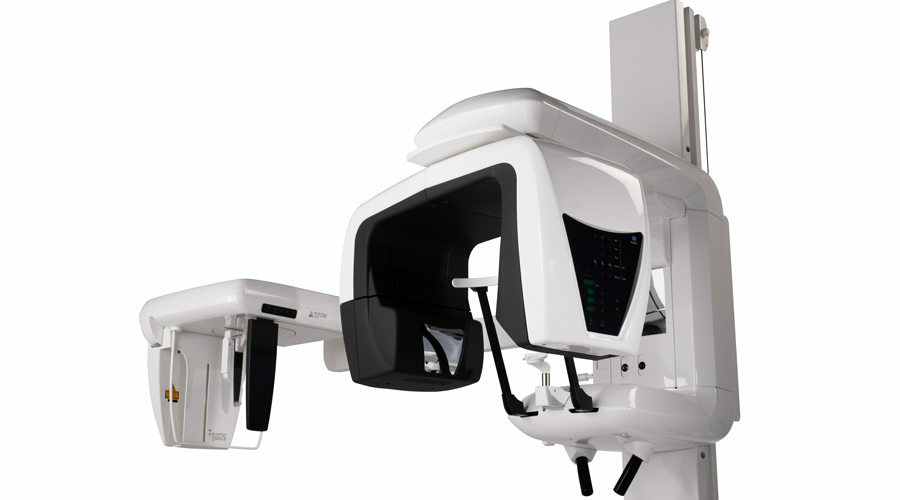
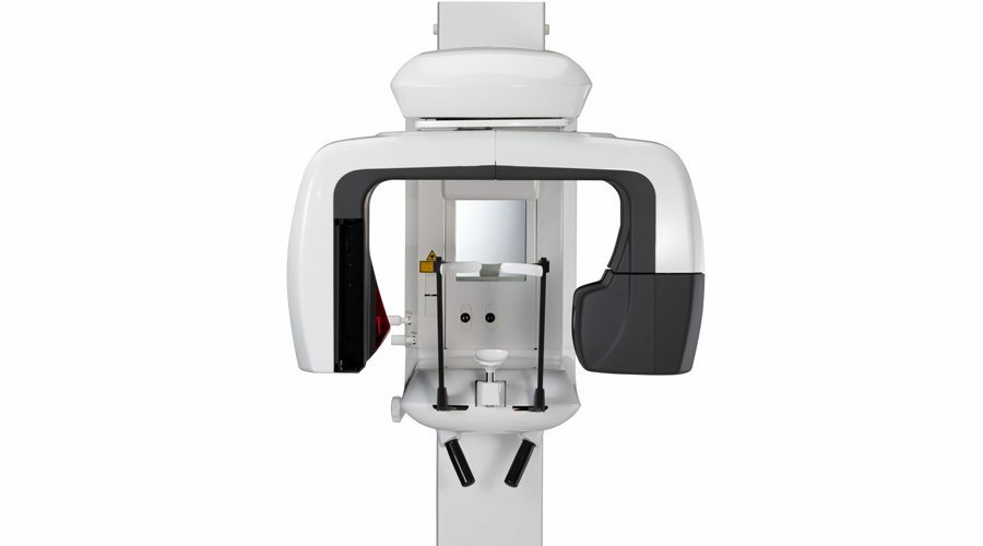
Veraviewepocs® 2D offers crystal clear panoramic and cephalometric images with low effective dose. Designed for versatility, the 2D model can be upgraded to 3D at a later date.

With fully automatic settings, Veraviewepocs 2D is easy to operate. This unit features a variety of options and specialized programs such as the Orthoradial Panoramic projection, which reduces the overlapping of neighboring teeth, and the Shadow Reduction Panoramic projection, which reduces obstructing shadows. Patient positioning is also easy with AF Automatic Positioning – a light beam sensor automatically positions the C-arm without requiring the patient to move.


NEW High Definition (HD) Cephalometric Update

Cephalometric image quality has recently been enhanced with Morita’s HD update. Advancements in image acquisition have resulted in X-rays with greater penetrating power. This new feature produces high definition, cephalometric images with amazing clarity and soft tissue display. It is now easier than ever before to plot key points for cephalometric measurements. This enhancement applies to this unit, as well as Veraviewepocs 3D, and Veraviewepocs 3De.

For accurate diagnosis and analysis, Veraviewepocs 2D offers high definition, refined imaging processing with multi-plane observation.


FEATURES

  • Completely upgradeable to the Veraviewepocs 3D model
  • 7.4 second panoramic; 4.9 second cephalometric
  • Super Fine Mode option for even higher resolution images
  • Continuous, automatic adjustment of kV and mA for excellent clarity
  • High quality images using both Digital Direct Automatic Exposure (DDAE) and Automatic Image Enhancer (AIE)
  • Easy patient positioning with AF automatic positioning, triple laser beams, and power assisted movement
  • Wide dynamic range of hard and soft tissue

Downloads

Related Products


High Resolution
In Fine High Speed Mode the pixel size is reduced by 25% compared to the previous model, so it produces superior images of a higher resolution. Super Fine Mode produces an even better image with increased resolution.


Multiple Projections
The distance from the X-ray tube to the patient is consistent, providing uniform magnification. The overlapping of neighboring teeth or the shadow of the opposing mandibular ramus is reduced, providing optimal results for jaw exposures.


Digital Direct Automatic Exposure (DDAE)
DDAE controls the X-ray tube voltage (kV) and current (mA) simultaneously by detecting X-rays passing through the patient. This improves the dynamic range, and along with Automatic Exposure (AE), results in exceptionally clear images with the best possible contrast and even density. The Automatic Exposure level can be adjusted to meet individual requirements.


Panoramic

Panoramic/Cephalometric