Please feel free to use our e-mail back service.
“Wherever possible and as long as the patient agrees to the resulting costs, I prefer using CBCT for questions regarding the osseous structures of the skull: The resolution is higher and radiation exposure lower than with a computer tomography. With CBCT, you always have a 3D dataset that can be viewed by the referring physician as well using the viewer that is provided free of charge.“
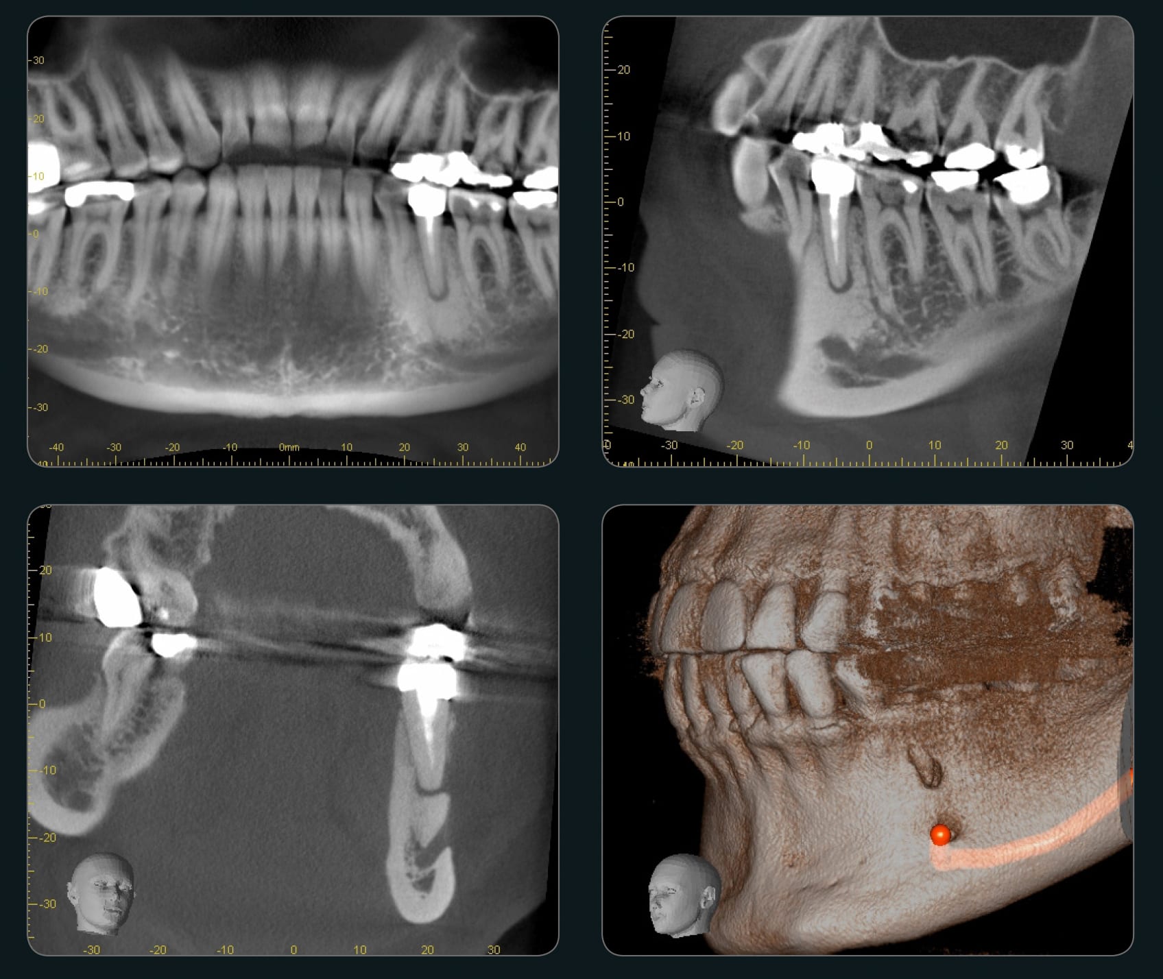
Case:
45-year old female patient with incomplete root filling 25 years ago.
Diagnosis:
Chronic periapical periodontitis
Findings:
The sclerosis in the bone and osseous defect in the alveolar process above the mental foramen are clearly visible. Three-dimensional reconstruction, in particular, clearly shows the close location to the mental foramen.
Therapy:
Root resection
Accuitomo Brechtelsbauer


