Please feel free to use our e-mail back service.
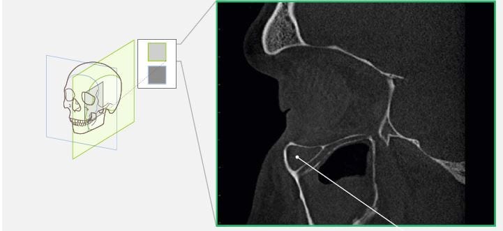
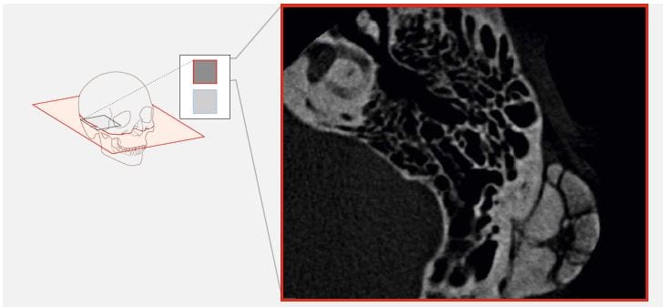
Case 1:
The patient reported a swelling behind the left ear, which exists since more than 10 years with very slow growing. Now he wished the operative extirpation so a cone beam tomography was done. The pictures show a osteoma of the temporal bone. History confi rmed this diagnosis.
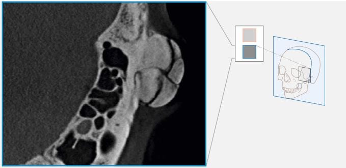
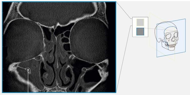
Case 2:
The patient reported about a chronic pain on the right maxillary sinus. Before the planned endonasal sinus surgery a cone beam tomography of nose and paranasal sinus were performed. A free bony course of the nervus infraorbitalis through the pathology of the right maxillary sinus could be seen. [This status was also seen in intraoperative endoscopic view]