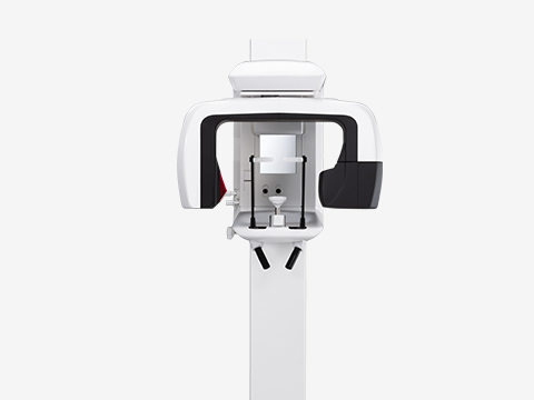
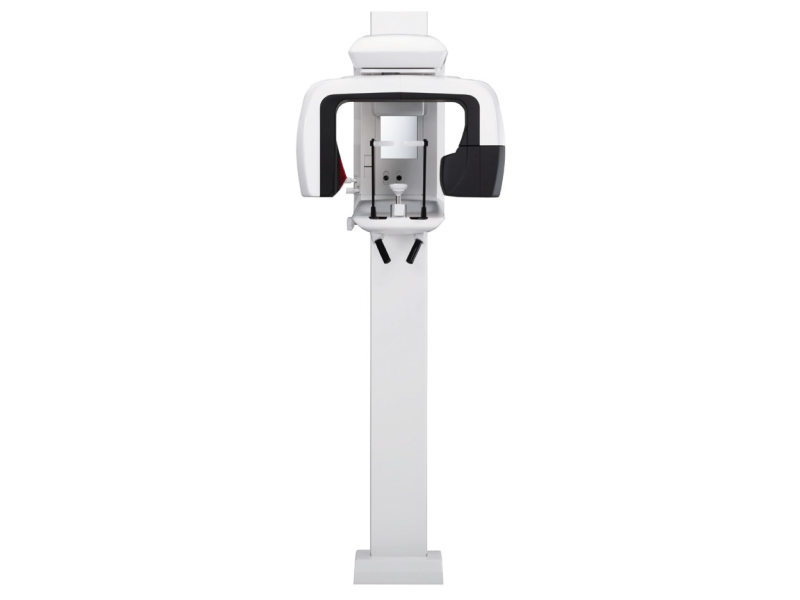
Veraviewepocs 2D

Veraviewepocs 2D High quality images for an accurate diagnosis

Fine or high speed mode

With high-speed to digital views… Read more

With high-speed to digital views:

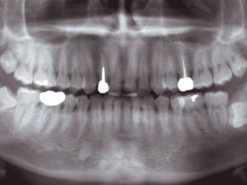
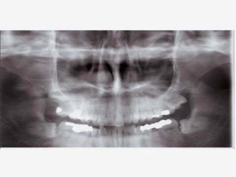
Digital Panoramic

Imaging exposure time only 7.4 seconds, thus only 1/4 of the radiation exposure incurred with traditional devices.


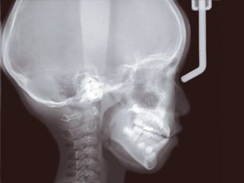
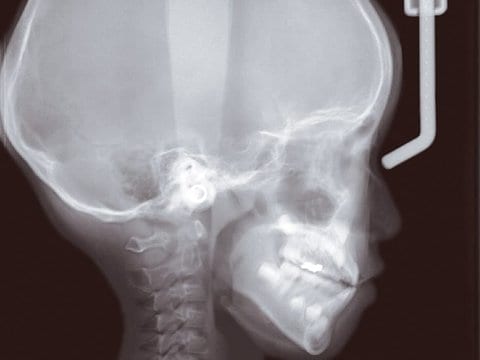
Digital cephalometric

Imaging exposure time approx. 4.9 seconds, thus only 1/10 of the radiation exposure incurred with traditional devices.


High quality images

Super High Quality Image – The Veraviewepocs has high resolution even in Fine High Speed Mode. The resulting image has high resolution, with superbdensity and contrast. Digital Direct AE… Read more

Super High Quality Image – The Veraviewepocs has high resolution even in Fine High Speed Mode. The resulting image has high resolution, with superbdensity and contrast. Digital Direct AE (automatic exposure) and Automatic Image Enhancer always obtains the optimal image.

Digital Direct Automatic Exposure (DDAE)

DDAE controls the X-ray tube voltage (kV) and current (mA) simultaneously by detecting X-rays passing through the patient. This improves the dynamic range, and, along with Automatic Exposure (AE), results in exceptionally clear images with the best possible contrast and even density. The automatic exposure level can be adjusted to meet your individual requirements.

Automatic Image Enhancer (AIE)

Automatic Image Enhancer enhances the details that can be observed in areas which are either extremely white or extremely black. DDAE and AIE perform a logarithmic conversion to produce the highest quality image possible.

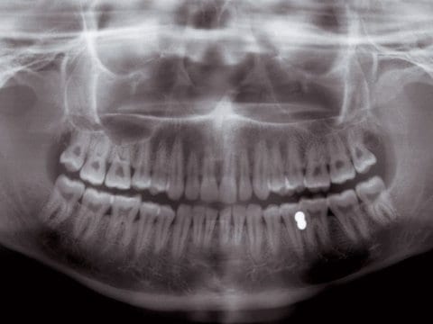
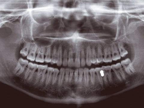
Conventional Image

Automatic Image Enhancer


High Resolution

Fine High Speed Mode: Pixel size is reduced to 25% compared with the former model, so it produces superior images of a higher resolution.

  • Pixel size Fine High Speed Mode: 144 µm
  • Pixel size Super Fine Mode: 96 µm
Simple and comfortable patient’s positioning

Easy, optimal patient positioning made possible with innovative technology… Read more

Easy, optimal patient positioning made possible with innovative technology.

AF Automatic Positioning

The Light beam sensor automatically positions the C-arm without the patient having to move. It then measures the distance to the patient's anterior teeth and AF automatically moves the C-arm into the optimum position. This creates images with a high degree of reproducibility.

Power-Assisted Movement: C-arm is Lined Up to the Patient

The electric motor of Veraviewepocs 2D enables convenient lift movement for smooth slow-starts and slow-stops. It is equipped with an automatic overload stop function for safety. In addition, the C-arm is lined up to the patient for easier patient positioning. Since the arm moves back and forth to line up with the patient, the patient does not have to move and can maintain a comfortable posture.

3 Laser Beams for Accurate Positioning

The patient‘s position is checked with the triple laser beams: the frankfurt plane beam, the sagittal plane beam, and the image layer beam for accurate positioning. The carbon temple stabilizing rods absorb almost no X-radiation and reduce the shadow of the rods in the image. The chin rest can be set at three different heights.

Wheelchair Support

The Veraviewepocs 2D offers a width of up to 480 mm to accommodate patients in wheelchairs. For patients with a wheel chair span greater than 480 mm, there is an optional wallmounted version available.


Versatile in mode and design

Multi-projections fit a variety of purposes. Consistent magnification is maintained throughout the image…
Read more

Multi-projections fit a variety of purposes. Consistent magnification is maintained throughout the image.
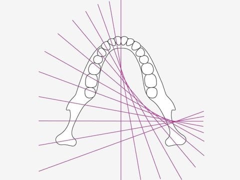
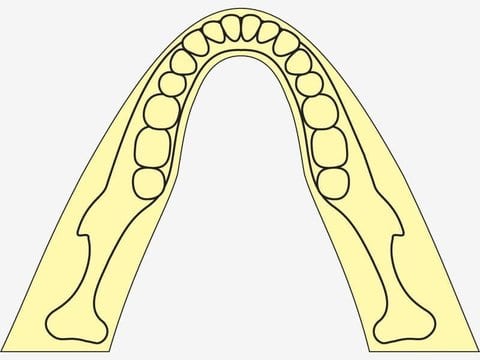
Standard Panoramic
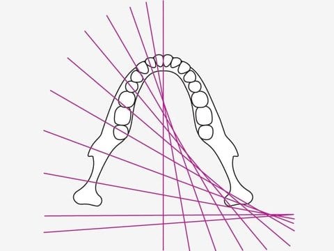
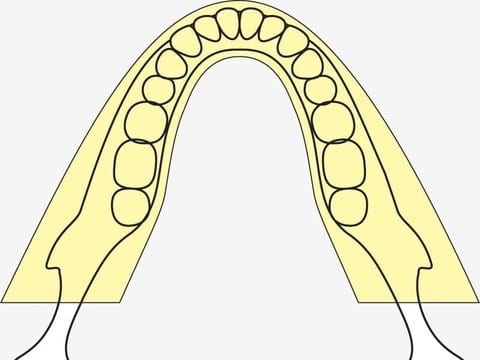
Shadow Reduction Panoramic
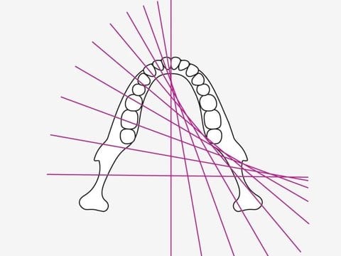
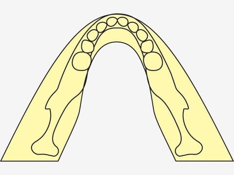
Orthoradial Panoramic

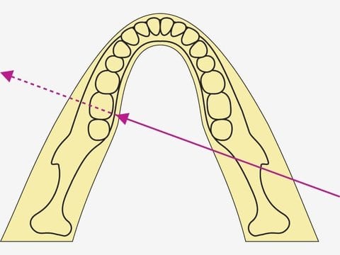
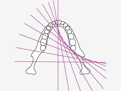
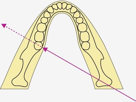
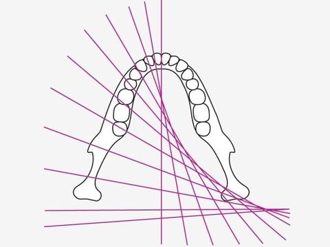
Distance from the X-ray tube to the patient is consistent, providing uniform magnification. In this way the overlapping of neighboring teeth or the shadow on the mandibular ramus is reduced, providing optimal results for jaw exposures. 

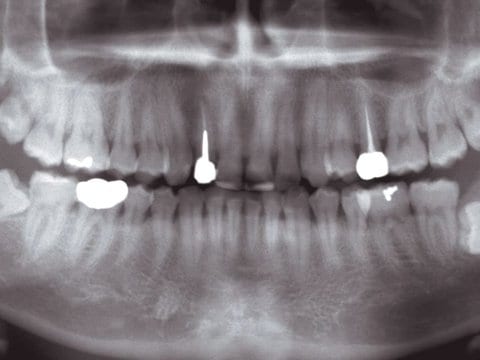
A) Digital Panoramic

Standard Panoramic, Mag.: 1.3 x constant

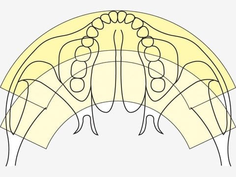
The thick / specially-designed image layer accommodates all the possible variations of dental arch shapes and sizes to produce extremely clear and sharp images.


Standard Panoramic, Mag.: 1.6 x constant

The X-ray image is enlarged by a factor of 1.6 – the best prerequisite for an even better diagnosis.


Pedodontic Panoramic, Mag.: 1.3 x constant (Mag.: 1.6 x is also available)

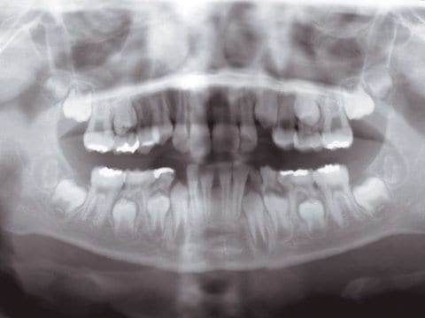
For children or people with small jaws. The arm‘s rotation range is reduced, and thus lessens the X-radiation. 


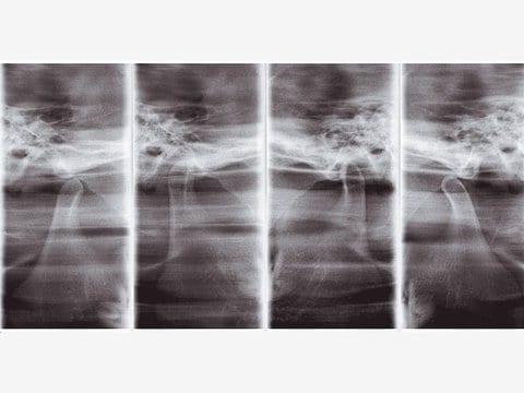
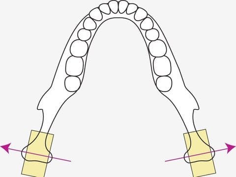
TMJ 4 Views, Mag.: 1.3 x constant

Sharp, clear images of the TMJ are produced by aligning the angle of X-ray penetration with the longitudinal axis of the mandibular condyle head.


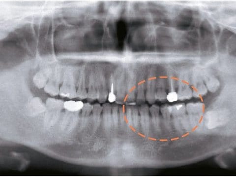
Orthoradial Panoramic, Mag.:1.3 x constant (Mag.: 1.6 x is also available)

The perpendicular projection of the X-ray reduces the amount of overlapping with emphasis on the maxillar bicuspid region.

Shadow Reduction Panoramic, Mag.:1.3 x constant (Mag.: 1.6 x is also available)

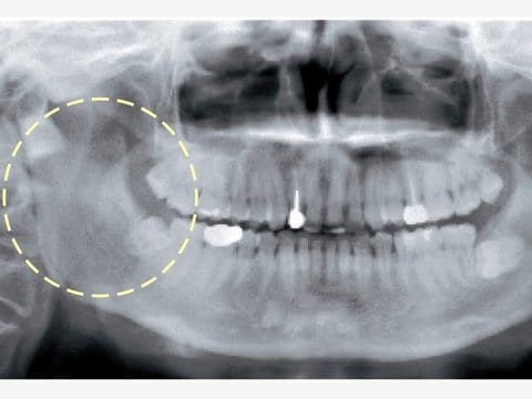
Produces images with less mandibular ramus shadow.

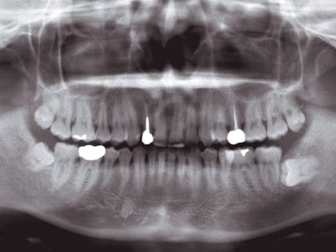
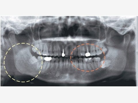
Please compare: Orthoradial Panoramic, shadow reduction panoramic, and standard panoramic are taken for the same patient.

Here would be changed the X-ray projection angle – and not the image layer orbit. In this way the overlapping of neighboring teeth or the shadow on the mandibular ramus is reduced. These images are good for diagnosis of dento-maxillo facial areas.

Standard Panoramic, Mag.: 1.3 x constant
  • Orthoradial panoramic for better observation of interproximal spaces
  • Shadow reduction panoramic for better observation of jaw
Maxillary Sinus Panoramic, posterior Mag.: 1.5 x constant

Clear Images of the Maxillary Sinus Region.


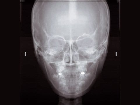
B) Digital Cephalometric

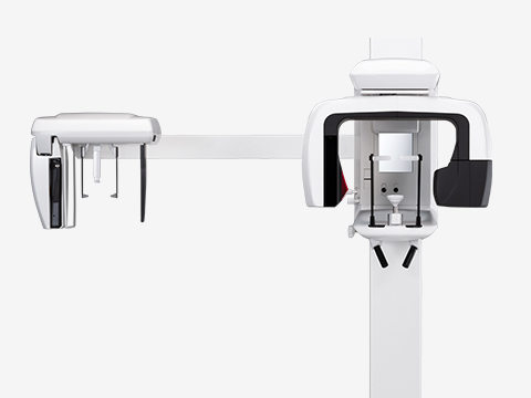
The following programs in the Ceph version are available:

  • posterior-anterior
  • lateral
  • Handwrist radiograph
Digital cephalometric – superfast, gentle and economical

The Veraviewepocs System offers high speed performance requiring only 4.9 seconds for a cephalometric scan. The speed helps ensure high quality images each and every time. For pediatric… Read more

The Veraviewepocs System offers high speed performance requiring only 4.9 seconds for a cephalometric scan. The speed helps ensure high quality images each and every time. For pediatric patients, the reduced scan time is especially helpful as repeat images due to patient movement is virtually eliminated.

  • High quality image with wide dynamic range
    You obtain far more information about hard and soft tissue – with just a single acquisition.
  • Imaging process can be completed within 20 seconds
  • A single digital cassette for panoramic and cephalometric
    A brand-new development: our special high-resolution CCD sensor with a height of 225 mm now makes digital cephalometric possible! Simply insert the new digital cassette and remove it again. One digital cassette can be used for both digital panoramic and digital cephalometric imaging.

  • Variable image processing capabilities
    The variable image processing technique generated optimum grayscale values – by offering different cassette running speeds for hard and soft tissue.
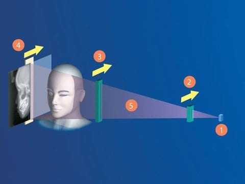
  1. Focal spot of X-ray tube
  2. Primary slit
  3. Secondary slit
  4. CCD sensor
  5. X-ray beam