
i-Dixel

i-Dixel Comprehensive image processing and diagnostics

Support for Multifaceted Diagnosis and Communication with Patients

The image diagnostic workstation i-Dixel is a software platform that enables processing and management of various types of images including CT images... Read more

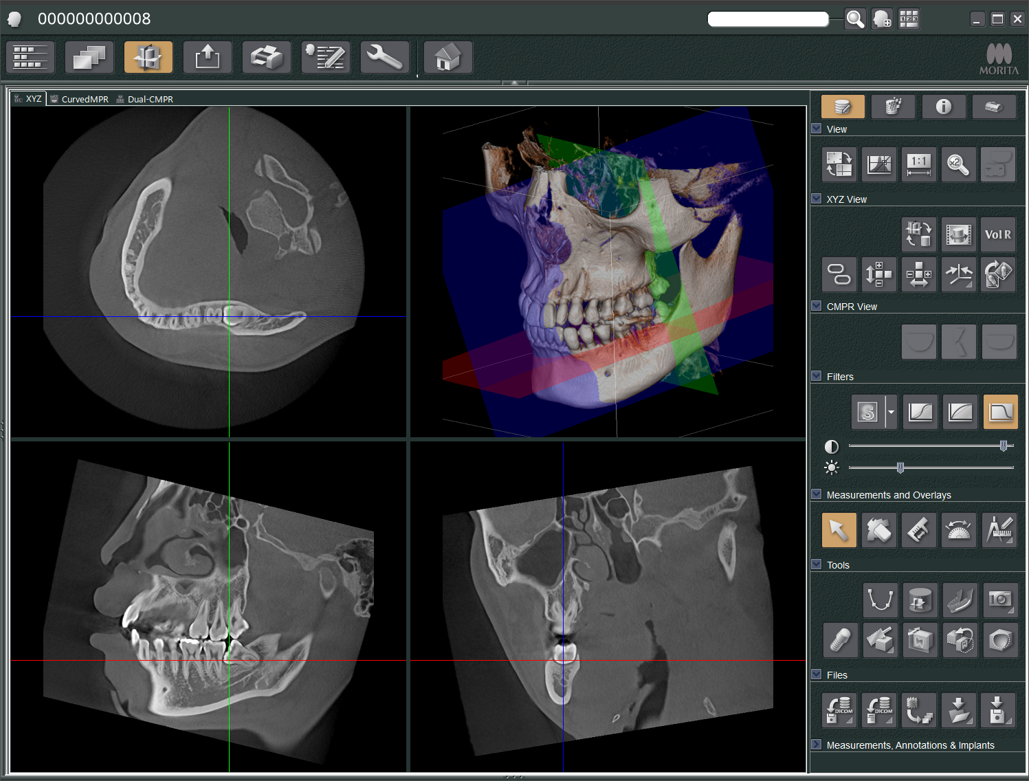
The image diagnostic workstation i-Dixel is a software platform that enables processing and management of various types of images including CT images, digital panoramic, cephalometric, dental X-ray images.
Its diverse functions extract a wealth of information from 2D and 3D images, supporting both diagnosis and patient explanations.

CT Airway Rendering Function (Optional)


CT


Panoramic


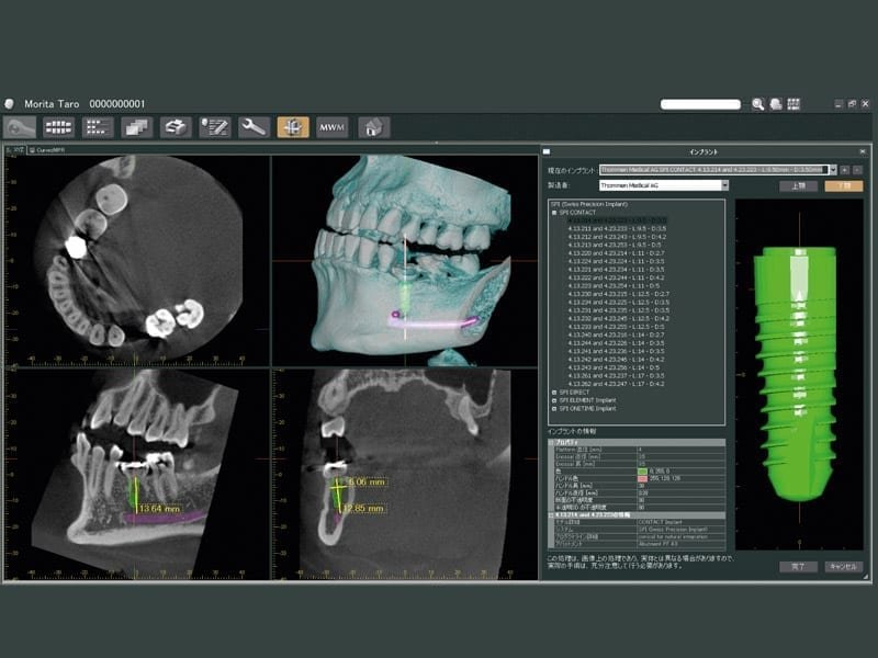
CT Implant Presentation Function


Cephalometric

Morita MAR

Reduce metal artifacts, improving image clarity

  • High-density objects can create artifacts in CBCT reconstructions, for example due to beam hardening.
  • These artifacts may obscure diagnostically relevant information.
  • They can also complicate digital dentistry applications in third-party software.

The MAR (Metal Artifact Reduction) function helps reduce metal artifacts, improving image clarity and supporting reliable downstream digital workflows. We are very pleased to already have received very good feedback from the endodontic community. Morita’s MAR is not AI generated and is based off the original input data and will produce an entirely different and unique reconstructed volume. The original volume is available for comparison.

MAR requires the use of H filters which generate pseudo-Hounsfield units so be sure to set your default reconstruction filters accordingly. From the nature of the machines, there are some conditions to be met to apply MAR. To learn if it is suitable for your Morita CBCT, you can contact your Morita sales representative.

Existing customers can update their existing licenses by purchasing an update script. Contact your local Morita representative for pricing information.

3D Viewer

By extracting the airway region* within a specified range on a CT image and marking and displaying it in 3D, the airway area can be... Read more

Airway Region on Sagittal Slice Image

Airway Region on Axial Slice Image

Airway Region on Volume Rendering

Airway Rendering Function (Optional)

By extracting the airway region* within a specified range on a CT image and marking and displaying it in 3D, the airway area can be visualized.
It also displays the volume of the extracted airway region and the cross-sectional area of the airway region on the currently viewed slice image.

*In this function, the “airway region” refers to the air-filled area within the range specified by users.

  • Compatible Equipment
  • 3D Accuitomo 
  • Veraview X800 / F150
3D for presentation and orientation

Keep yourself oriented and help present treatment plans to your patients with 3D volume rendering. i-Dixel makes it easy and provides you multiple ways to view your scan from any angle... Read more

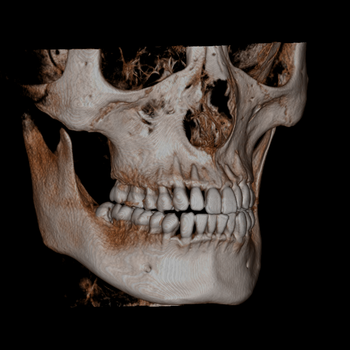
Keep yourself oriented and help present treatement plans to your patients with 3D volume rendering. i-Dixel makes it easy and provides you multiple ways to view your scan from any angle imaginable. From bone structure to calcifications, i-Dixel's volume rendering helps you get a sense of the extent of bone loss and allows you to see minute changes in density that may affect your treatement plans.

The volume rendered image is can be linked to the slices so you are always oriented and this position is always updated in realtime.
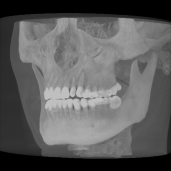
Maximum Intensity Projection


Mandibular Canal Depiction

Mark the mandibular canal in 3D to ensure visibility in all slices and all volume renderings... Read more

Mark the mandibular canal in 3D to ensure visibility in all slices and all volume renderings. i-Dixel makes it easy to see the mandibular canal and helps ensure implants are placed safely. Buccal and lingual position becomes immediately clear from any view.

Implant Presentation Preparation

This feature allows you to specify the length and diameter of implant
fixtures and paste them into the image. This presentation is easily understood by patients and helps with case… Read more

This feature allows you to specify the length and diameter of implant fixtures and paste them into the image. This presentation is easily understood by patients and helps with case acceptance.

Stand-alone viewers for data sharing

i-Dixel provides a fully-featured data viewer that can be exported and shared with referring doctors or colleagues... Read more

i-Dixel provides a fully-featured data viewer that can be exported and shared with referring doctors or colleagues. Your annotations are secured and cannot be changed once exported ensuring your notes are never changed.

The viewer can run on any Windows PC and displays the CT data exactly as shown in i-Dixel. All features and annotations are avaialalbe for use with the exception of one: saving.

DICOM 3.0 Compatible

i-Dixel Supports the following DICOM Service Classes… Read more

i-Dixel Supports the following DICOM Service Classes:

  1. Modality worklist management service class
  2. Storage service class
  3. Modality performed procedure step service class
  4. Print management service class

Standardization and Information Exchange