9 fields of view for flexible scanning from local to large areas
The 3D Accuitomo is equipped with 9 FOVs (fields of view) that allow flexibility when scanning patients with a variety of diagnostic needs and clinical indications, from a large area (Ø170 × H 120 mm) that covers the maxillofacial region to a local area (Ø40 × H 40 mm).
Reducing exposure dose is possible by selecting the most suitable FOV.
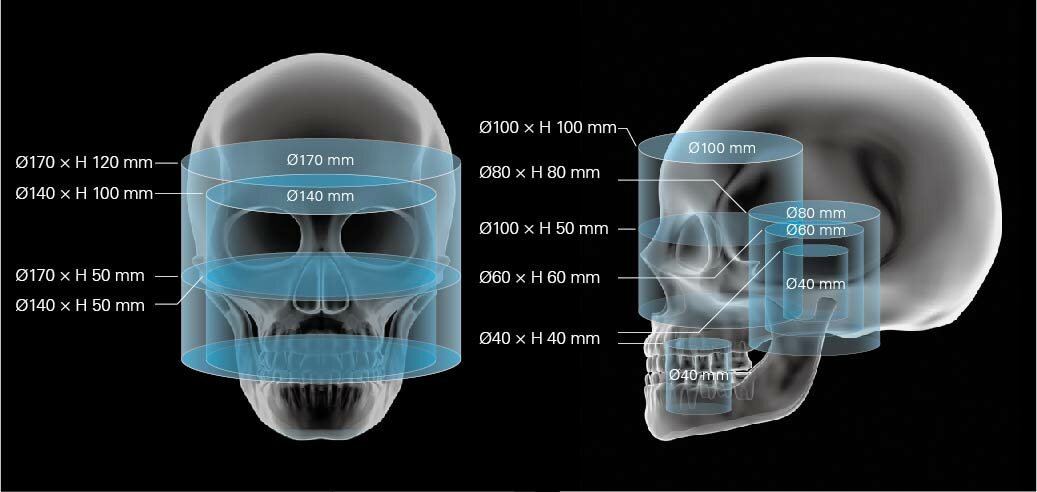
Fields of View
| FOV |
Voxel Size |
| Ø 40 x H 40 mm |
80μm |
| Ø 60 x H 80 mm |
100μm |
| Ø 80 x H 80 mm |
125μm |
| Ø 100 x H 50 mm |
160μm |
| Ø 100 x H 100 mm |
| Ø 140 x H 50 mm |
200μm |
| Ø 140 x H 100 mm |
| Ø 170 x H 50 mm |
250μm |
| Ø 170 x H 120 mm |
High resolution even at large FOVs
The minimum voxel size can be selected from 80 μm, 100 μm, 125 μm, 200 μm, or 250 μm depending on your diagnostic needs and clinical indications.
The 3D Accuitomo is able to provide high resolution with less distortion, even at large FOVs.
The FOV can be offset so that even the temporal bone region can be positioned at the center of the FOV. This results in well-focused, high resolution image
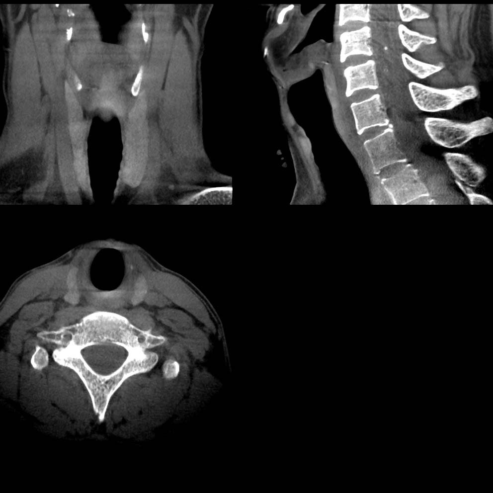 Ø 140 x H 100 mm (200μm)
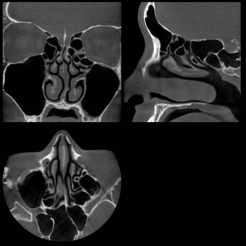 Ø 100 x H 100 mm (160μm)
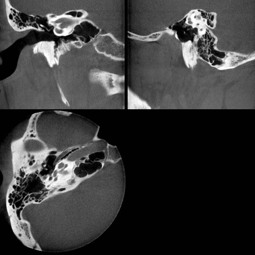 Ø 60 x H 60 mm (100μm)
Zoom reconstruction from original data
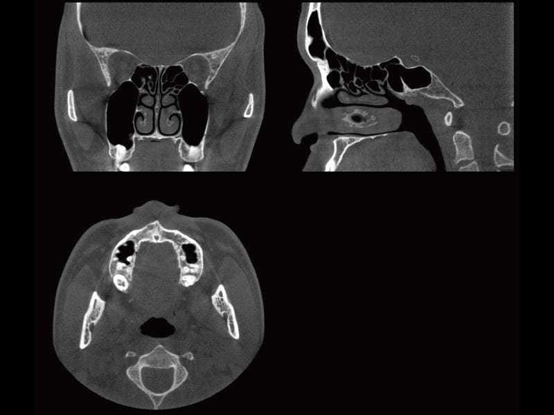
]The 3D Accuitomo is equipped with a unique zoom reconstruction function allowing you to zoom in and reconstruct a new volume from the original scan, without the need for additional acquisitions. The new volume can be reconstructed with a resolution of up to 80μm improving diagnostic accuracy with no additional X-ray exposure to the patient.
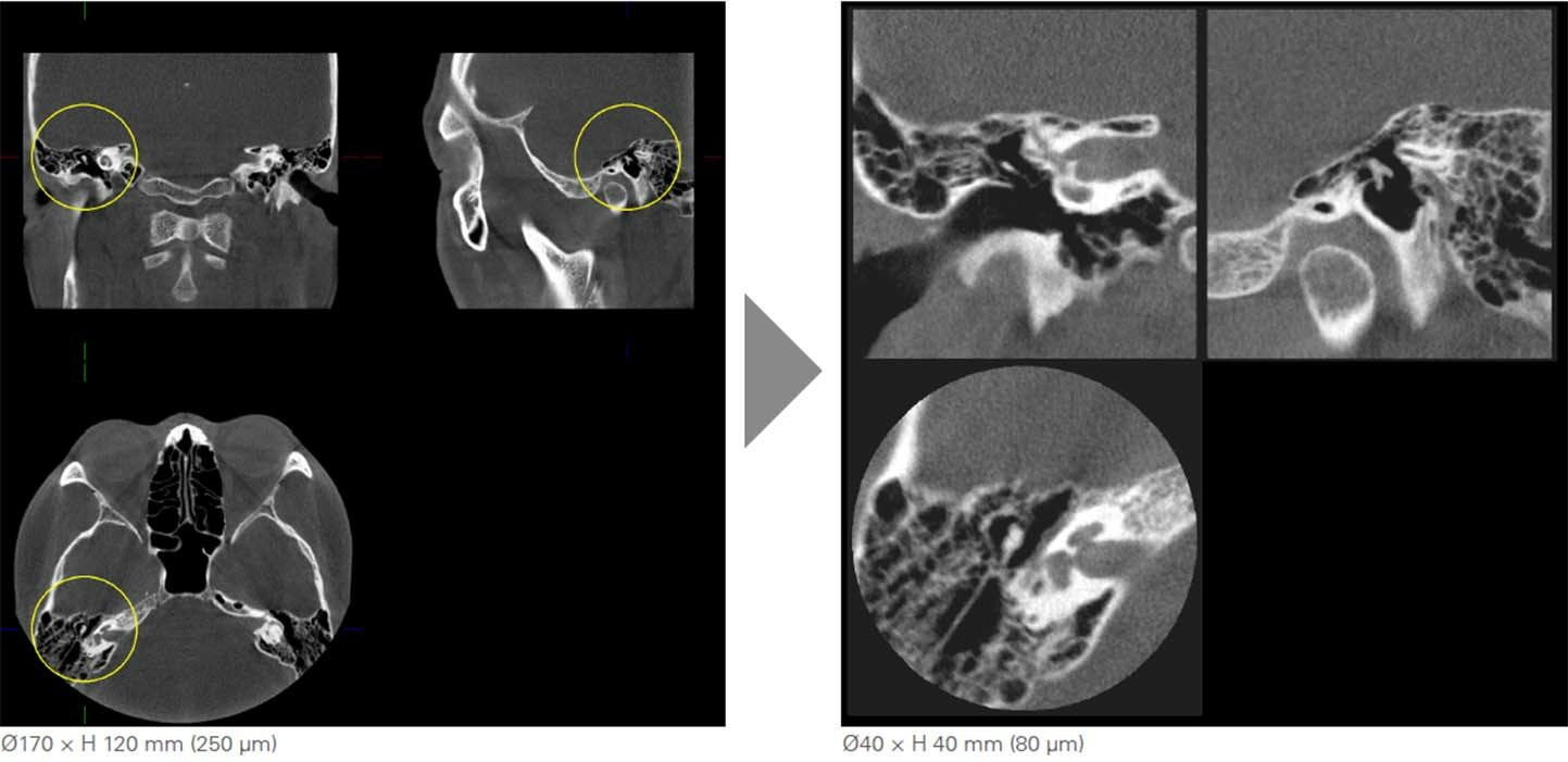
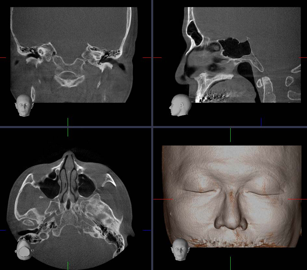 Ø170×H 120 mm (250 μm)
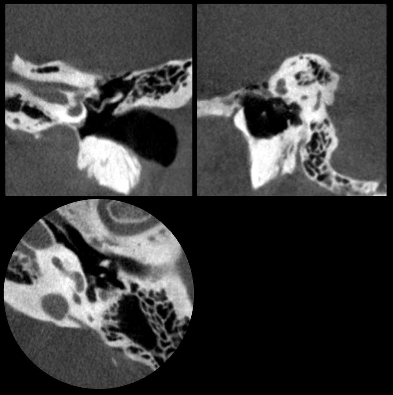 Ø40×H 40mm (80 μm)
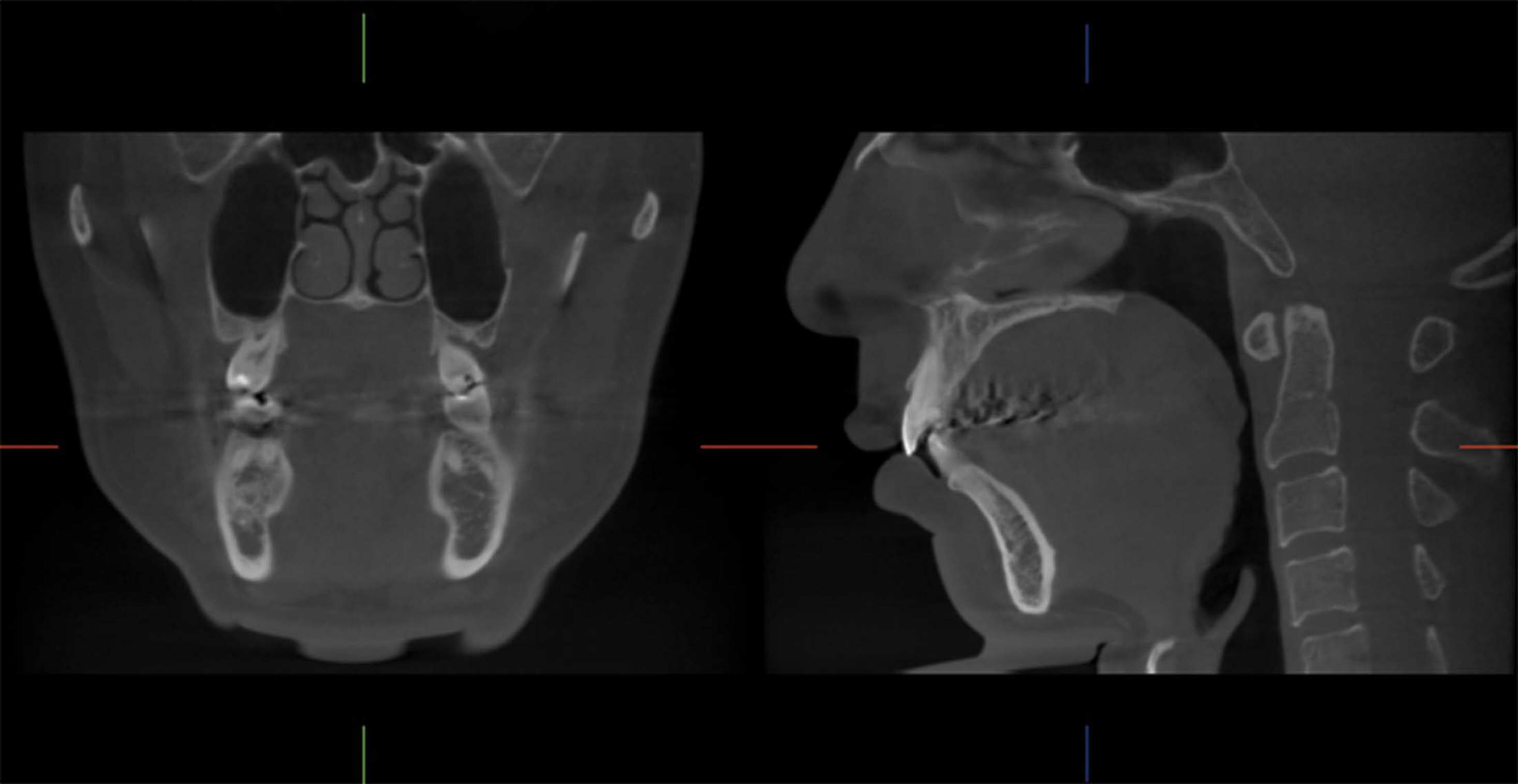 Ø170×H 120mm (250 μm)
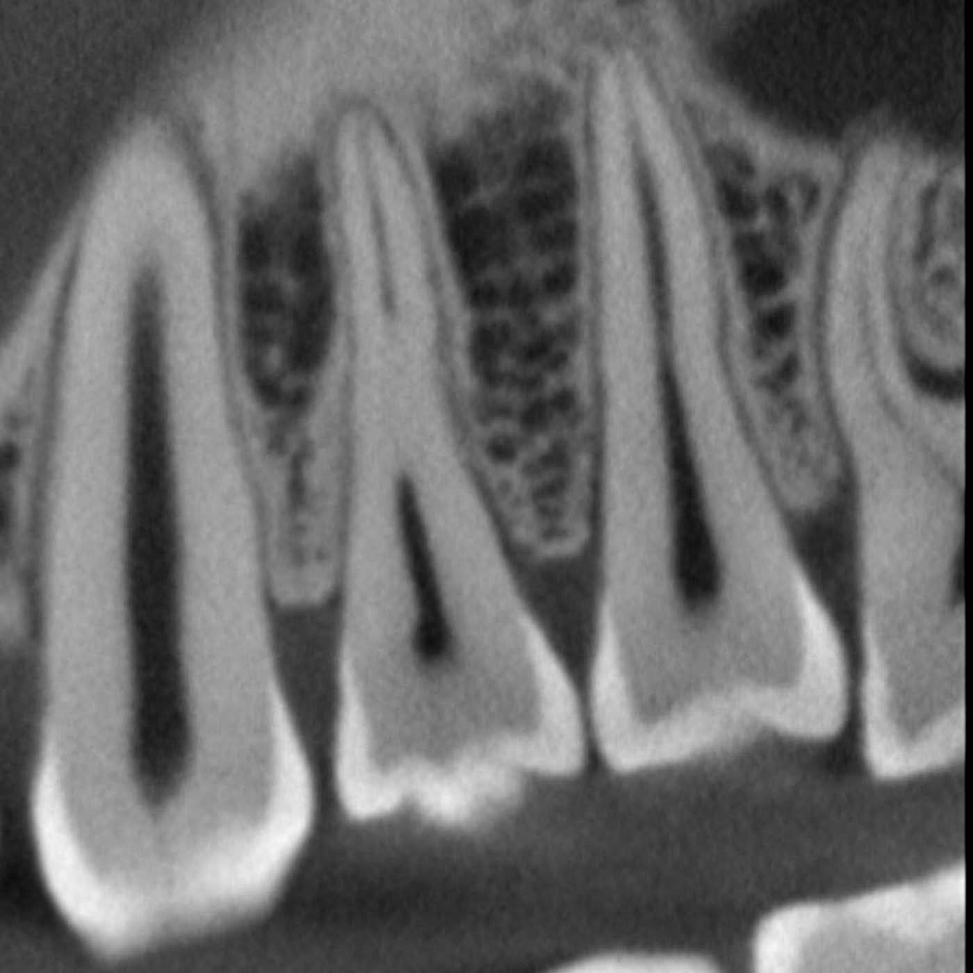 High Resolution Mode (80 μm)


 Ø 140 x H 100 mm (200μm)
 Ø 100 x H 100 mm (160μm)
 Ø 60 x H 60 mm (100μm)
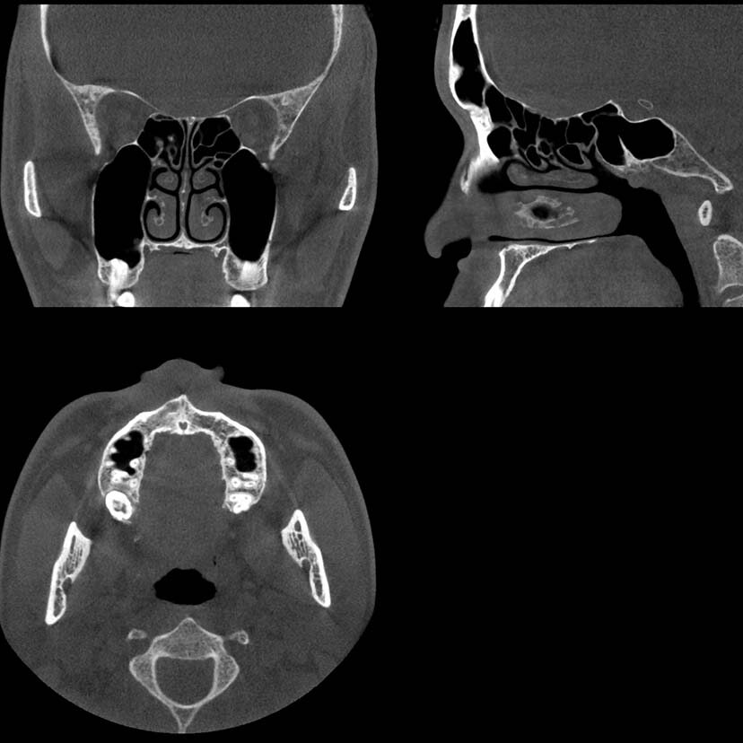 Standard Mode Ø170 mm × H 120 mm
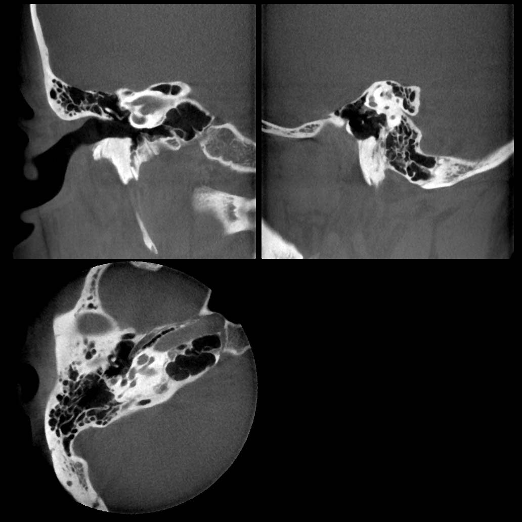 High Fidelity Mode Ø80 mm × H 80 mm
 Mastoidectomy Mode (neural tubes drawing and CT volume removing)
 Pseudo Rigid Scope Mode (perspective projection)


