Please fill out this contact form. We will come back to you soon.
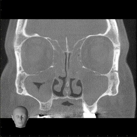
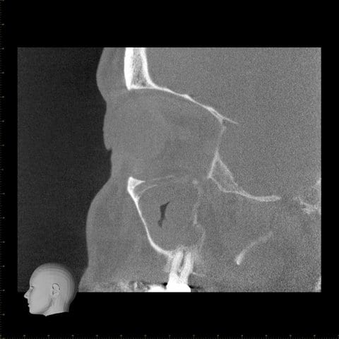
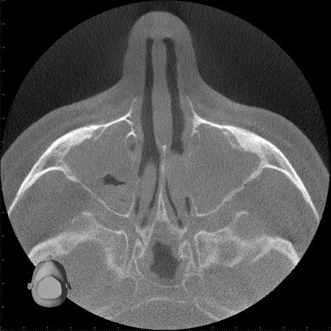
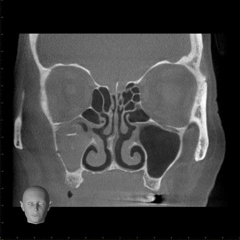
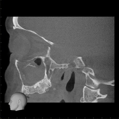
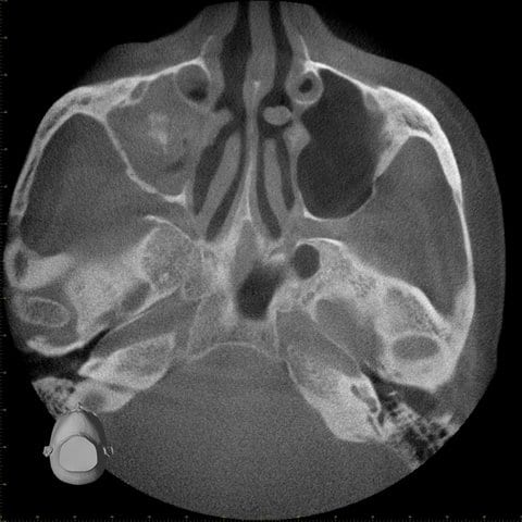
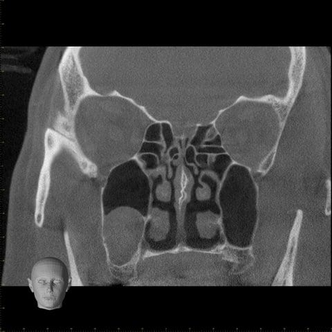
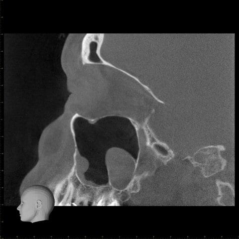
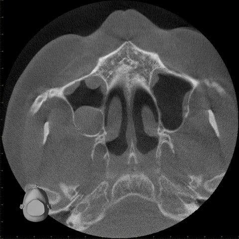
„The three-dimensional representation of the paranasal sinuses, middle ear, petrous temporal bones and the upper respiratory tracts, lets us answer diagnostic questions quickly, accurately and using only one system. Moreover, we can plan operations exactly. The examination is pleasant for patients because they can remain in a sitting position; and despite maximum resolution, the radiation exposure is significantly lower than in customary CTs. Dentists and oral surgeons can also benefit from the information obtained. Accordingly, interdisciplinary cooperation also provides economic benefits.“

