Please feel free to use our e-mail back service.
Diagnostic quality always has been a primary concern in connection with therapy and treatment planning. Many practices still send their patients to a radiologist for a CT when they require 3D images, even though cone-beam computed tomography is a great way to provide this service in an ENT practice. In addition to the advantages of continuous workflow and retention of the fees in your practice, you will spare your patients unnecessary radiation doses as compared to the CTs extensively being used in the market.
- 1. Our solution
- 2. Benefits of the technology
- 3. Technology
- 4. Technical qualification
- 5. Indications
-
Our solution

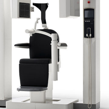
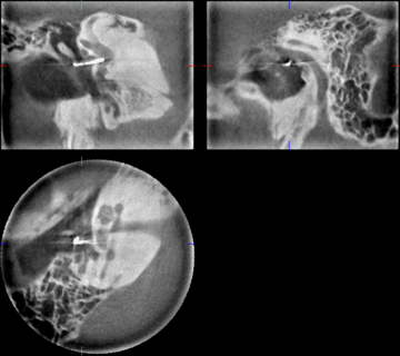
The CBCT scanner, 3D Accuitomo 170, is an ideal imaging system for your radiology practice. It provides detailed views of very fine anatomical structures in the areas of the head and neck, such as the temporal bone, paranasal sinus, eye sockets, jaw and skull base. Consequently, it can be used for ENT, plastic surgery and all dental indications.
-
Benefits of the technology

Three-dimensional imaging has one significant advantage: it can visualize anatomical structures in high definition. Hence, the doctor can obtain more data from the information because the true location of anatomical structures can only be discerned with a three-dimensional visualization. Accordingly, it helps make for safer diagnoses.
-
Technology

CBCT works on the basis of a cone-/pyramidal-shaped X-ray beam: for this purpose, a flat panel detector and an X-ray source are mounted on opposite sides of a rotating arm. The physician positions the patient in the isocenter, and the C-arm rotates at least 180° during the scanning process. While the scanner is rotating around the patient, the projection of a cylindrical volume is obtained at defined view angles with the cone-shaped X-ray beam.
-
Technical qualification
Why is a Technical Qualification Course necessary?
A Technical Qualification Course has to be completed to operate a CBCT system and to evaluate three-dimensional X-rays because only then will the physician be able to take CBCT X-rays and evaluate the images competently.
What does such a course entail?
The first module conveys technical instruction, "Taking skull X-rays for ENT indications", followed by technical instruction and proficiency modules in CBCT diagnostics. The CBCT Technical Qualification Course is held on two days respectively; however, there should be an interval of at least three months between the two course dates. Following regular attendance and successful completion of the final exam, the medical association will grant you the qualification to operate a CBCT system and evaluate three-dimensional X-rays.
-
Indications
Diseases of the paranasal sinus
- Acute/chronic ethmoidal sinusitis
- Acute/chronic frontal sinusitis
- Acute/chronic sphenoidal sinusitis
- Vacuum cephalgia
- Osseous defects in the paranasal sinus
- Planning surgical procedures in the paranasal sinus
Fractures
- Midfacial fractures
- Blow-out fractures
- Zygomatic arch fractures
- Nasal bone fractures
- Petrous bone fractures
Ear
- Primary and secondary cholesteatoma
- Mastoiditis (especially when affecting children)
- Stapes luxations (post-op)
- Cochlear implants (post-op)
- Semicircular canal dehiscence
- Malformations of the middle ear
Throat
- Foreign-body localization (if roentgenopaque)
- Swallowing diagnostics
- Sialoliths (if roentgenopaque)


