Please fill out this contact form. We will come back to you soon.
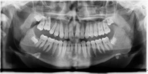
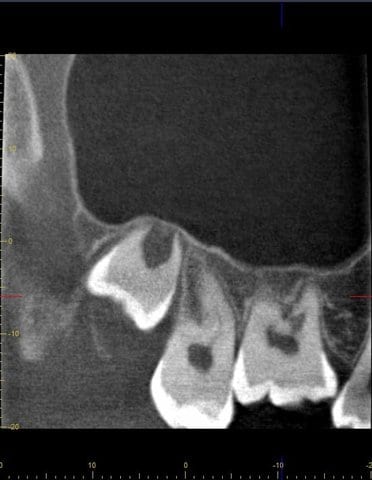
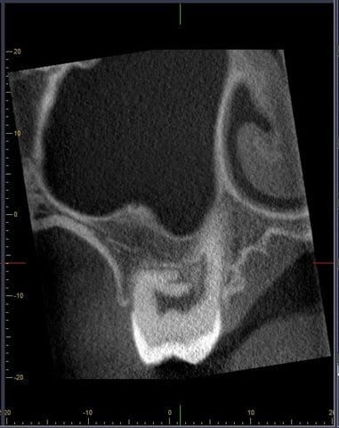
“The case presented here illustrates very clearly the advantages of 3D diagnosis in comparison to conventional 2D X-rays. A conclusive appraisal of the actual extent of local findings and optimum planning of the resulting treatment was only possible as a result of the CBCT scan.“
Case:
The patient was referred by his orthodontist to discuss possible therapies for tooth 16.
Findings:
Infraposition of tooth 16 with, as compared to the neighboring teeth, higher resonance and negative vitality test.
Diagnosis:
Presumption diagnosis: Ankylosis of tooth 16. Diagnosis (after CBCT): Vascularization of bone tissue up into the pulp in the furcation region.
Therapy:
CBCT to verify the presumption diagnosis. Extraction of tooth 16 and referral back to the oral surgeon for mesialization of teeth 17 and 18.


