Please feel free to use our e-mail back service.
Case:
Patient Female, 19 years old
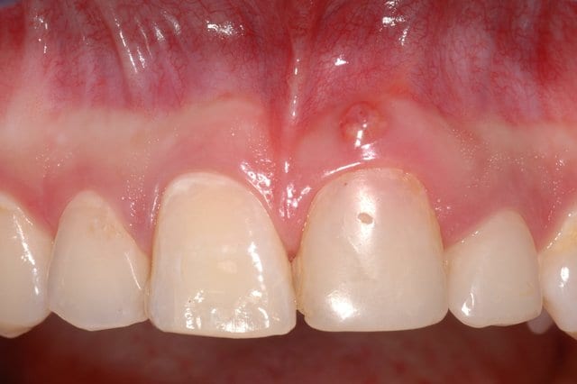
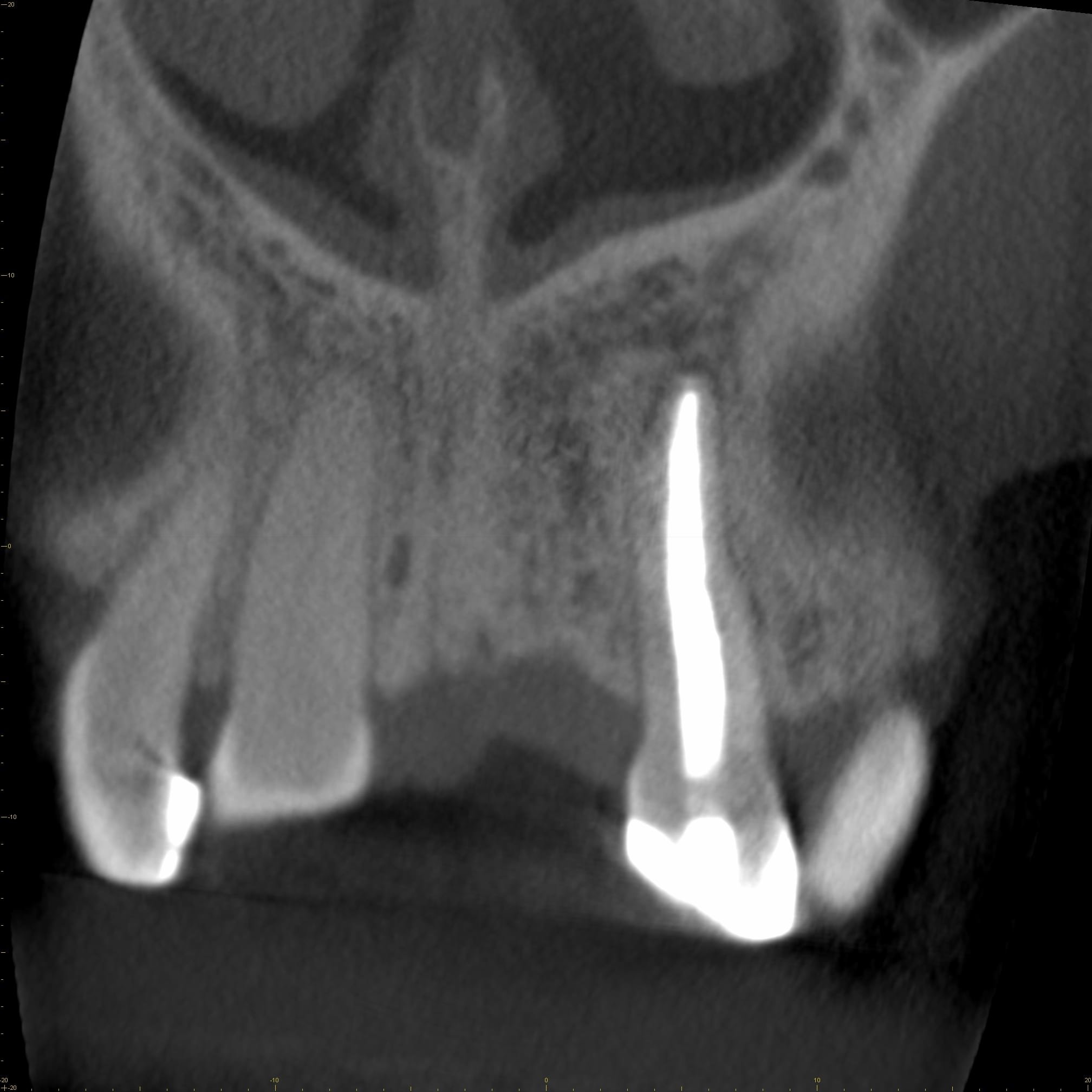
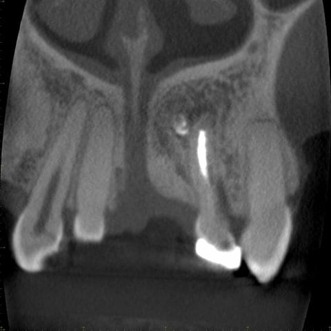
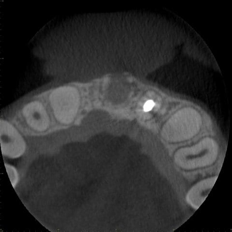
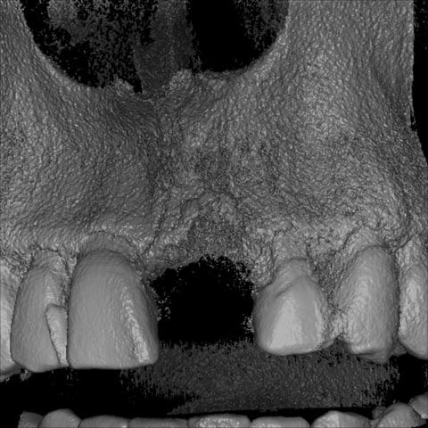
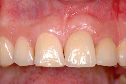
Initial clinical presentation of a 19-year old female patient referred for dental implant treatment planning. The patient had an avulsion of the maxillary left central incisor with 8 years. The tooth was replanted using a titanium post. Since a few months, the patient had noticed a buccal fistula and growing „pinkish“ discoloration of the tooth.
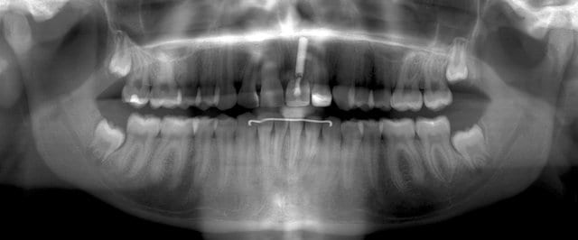
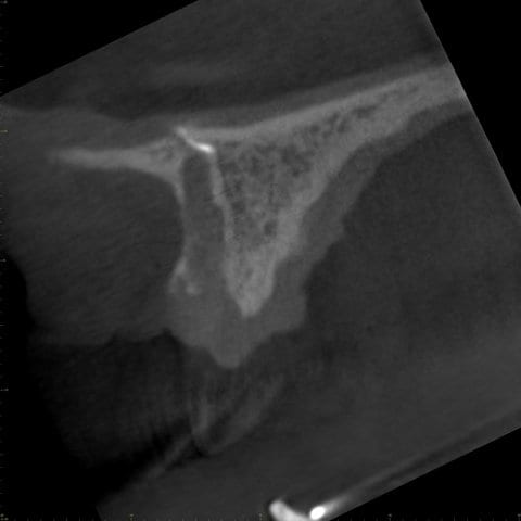
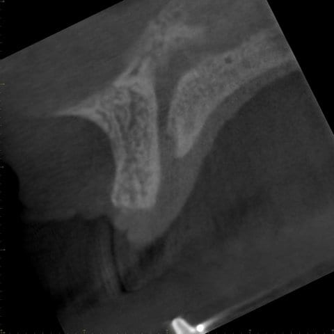
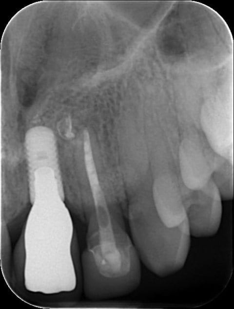
Panoramic view of the patient exhibiting the replanted maxillary left central incisor. A lytic, hypodense region is visible between the crown of the respective tooth and the titanium post. The root of the central incisor is not discernible – and seems to have been resorbed in most parts. Additional findings include non-erupted third molars in the maxilla and mandible.
Radiographic (A) and clinical (B) presentation two years after dental implant insertion in the region of the left maxillary central incisor.

