Please feel free to use our e-mail back service.
ENT specialists rely on high resolution, contrast rich images to properly diagnose problems in the head and neck region. Many practices send their patients to a radiologist for a CT or CAT scan when they require 3D images, even though cone beam computed tomography (CBCT) is a great way to provide this service in-house. In addition to the advantages of continuous workflow and retention of the imaging fees, you will spare your patients unnecessary radiation exposure as compared to CT scans still extensively being used in the market.
-
High Clarity

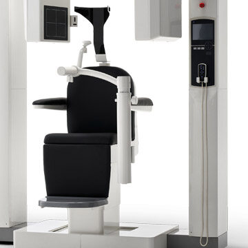
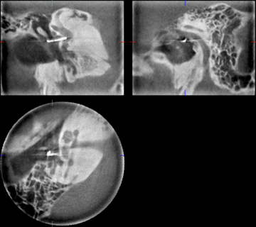
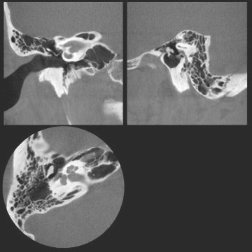
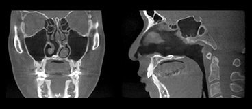
The 3D Accuitomo 170 offers a minute voxel size of just 80 μm (micrometers). This super-fine voxel displays an amazing level of clarity enabling a comprehensive examination. The imaging area is cylindrical and the maximum size is 170 mm in diameter by 120 mm in height, which covers the majority of the head and neck region, suitable for specialized medical applications in otorhinolaryngology and the maxillofacial field.
-
Low Dose

Patients benefit from a CBCT scan, compared to a conventional CT scan, because the radiation dose is significantly lower. For a CT image, the radiation source scans the region of the body that is to be examined in slices of 0.5 to 3.0mm; CBCT scans the entire section in one single rotation. As a result, the dosage is reduced by up to 80 percent as compared to a standard CT X-ray*.
* 1mm slice thickness, 1.5mm pitch, 120 mAs/rotation, 87mm scan height
-
How it works

CBCT works on the basis of a cone-/pyramidal-shaped X-ray beam: For this purpose, a flat panel detector and an X-ray source are mounted on opposite sides of a rotating arm. The physician positions the patient in the isocenter, and the C-arm rotates at least 180° during the scanning process. While the scanner is rotating around the patient, the projection of a cylindrical volume is obtained at defined view angles with the cone-shaped X-ray beam.
-
Indications

Otology
- Cholesteatoma
- Otosclerosis
- Mastoiditis
- Ossicular chain form
- Cochlear implant (CI)
- Semicircular canal dehiscence syndrome
- Congenital abnormalities of the middle ear
- Patulous Eustachian tube (PET)
- Temporal bone fracture
Rhinology
- Acute/Chronic Sinusitis
- Vacuum cephalgia
- Osseous defects in the paranasal sinus
- Planning surgical procedures in the paranasal sinus
- Image acquisition of IGS for FESS
Skull Base Surgery
- Endoscopic transnasal approach for skull base surgery with image guidance system
Laryngology
- Diagnosis of foreign body larynx (in the case of X-ray opaque)
- Ptyalolith (in the case of X-ray opaque)
- Form of epiglottis
Fractures
- Midfacial fractures
- Blow-out fractures
- Zygomatic arch fractures
- Nasal bone fractures
- Petrous bone fractures


