Please feel free to use our e-mail back service.
Morita is equipped to assist the periodontal office from diagnosis through treatment. The periodontal product line includes CBCT units with world-renowned clarity for evaluation, an Er:YAG laser and surgery blades for treatment, as well as bone augmentation material for implant preparation when the tooth cannot be saved.
Periodontal Workflow
-
Diagnosis & Planning

For periodontal diagnosis and evaluation, Morita offers CBCT units that deliver high resolution images with world-renowned clarity. These images provide a clear observation of the periodontal pocket, the periodontal membrane, and the alveolar bone allowing for complete evaluation of periodontics defects, implant failure (including peri-implantitis) and treatment planning.
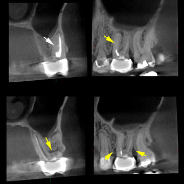
Case description: This 46 year-old female patient received endodontic treatment in tooth #14 months prior. The patient remained symptomatic after endodontic treatment. Clinical examination and periapical radiograph were unremarkable. On the periapical radiograph taken, the alveolar bone, lamina dura, periodontal ligament space and sinus floor appeared normal. The 3D Accuitomo scan illustrated two significant findings. The presence of an unfilled MB2 canal is seen in the MB root of #14 (white arrow). Importantly, widening of the periodontal ligament space consistent with persistent periapical disease is seen at the apex of the MB root. Additionally, vertical three-wall periodontal defects at the mesial and distal surfaces of #14 and loss of furcational periodontal bone loss were shown to be present (yellow arrows).
Case Courtesy of Sotirios Tetradis, DDS, PHD, Professor, Chair of the Section of Oral and Maxillofacial Radiology, UCLA School of Dentistry
-
Treatment

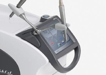
The Er:YAG laser eliminates vibration and is attracting considerable attention as a new treatment method. The wavelength of the Er:YAG laser is most suitable for dental treatment because it is readily absorbed by water. Therefore, it efficiently vaporizes human tissue that has high water content.
Wide Variety of Applications
The wide array of tip options enables this laser to perform both hard and soft tissue procedures.
Hard Tissue Treatment
(ablation, vaporization)
Class I, II, III, IV and V cavity preparation
Caries removalPeriodontal Treatment
(incision, excision, vaporization, ablation and coagulation)
Removal of subgingival calculi
Laser soft tissue curettageSoft Tissue Treatment
(incision, excision, vaporization, ablation and coagulation)
Gingival incision and excision
Hemostatis and coagulation
Frenectomy and frenotomy -
Observation

After surgery or treatment, Veraviewepocs 3D R100 and 3D Accuitomo 170 are excellent tools for CBCT observation. Clinicians can be comfortable taking follow-up x-rays as necessary. In additional to high resolution images, Morita units offer several patient protection features including a dose reduction mode. A typical 40 x 40 mm scan emits less dose than two standard panoramic X-rays.
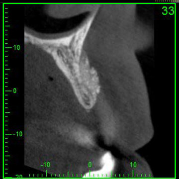
The clinical image to the left shows a CBCT evaluation of an implant site 6 months after guided bone regeneration.
Case image courtesty of Erika Benavides, DDS, PhD, Clinical Associate Professor and Hector Rios, DDS, PhD, Assistant Professor - Department of Periodontics and Oral Medicine, University of Michigan School of Dentistry.
-
Customer Statements
I used the large Foundation and was able to place an implant with great bone density.
-
Wilfred J. St. Cyr, DDS Catonsville, Maryland, USA
-


