Figure 1
Initial clinical presentation of a 19-year old female patient referred for dental implant treatment planning
The patient had an avulsion of the maxillary left central incisor with 8 years. The tooth was replanted using a titanium post. Since a few months, the patient had noticed a buccal fistula and growing „pinkish“ discoloration of the tooth.
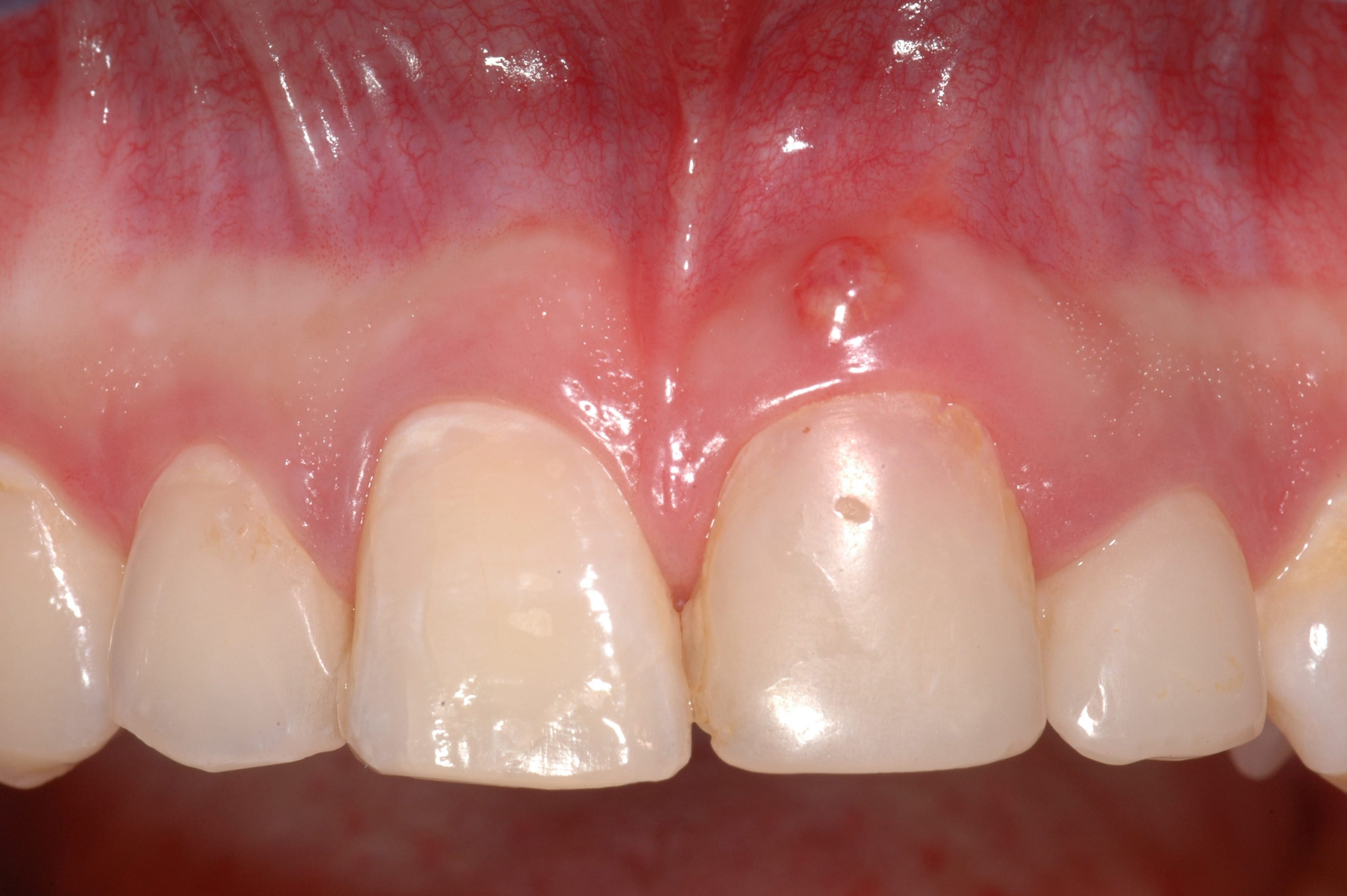
Figure 2
Panoramic view of the patient exhibiting the replanted maxillary left central incisor. A lytic, hypodense region is visible between the crown of the respective tooth and the titanium post. The root of the central incisor is not discernible – and seems to have been resorbed in most parts. Additional findings include non-erupted third molars in the maxilla and mandible.
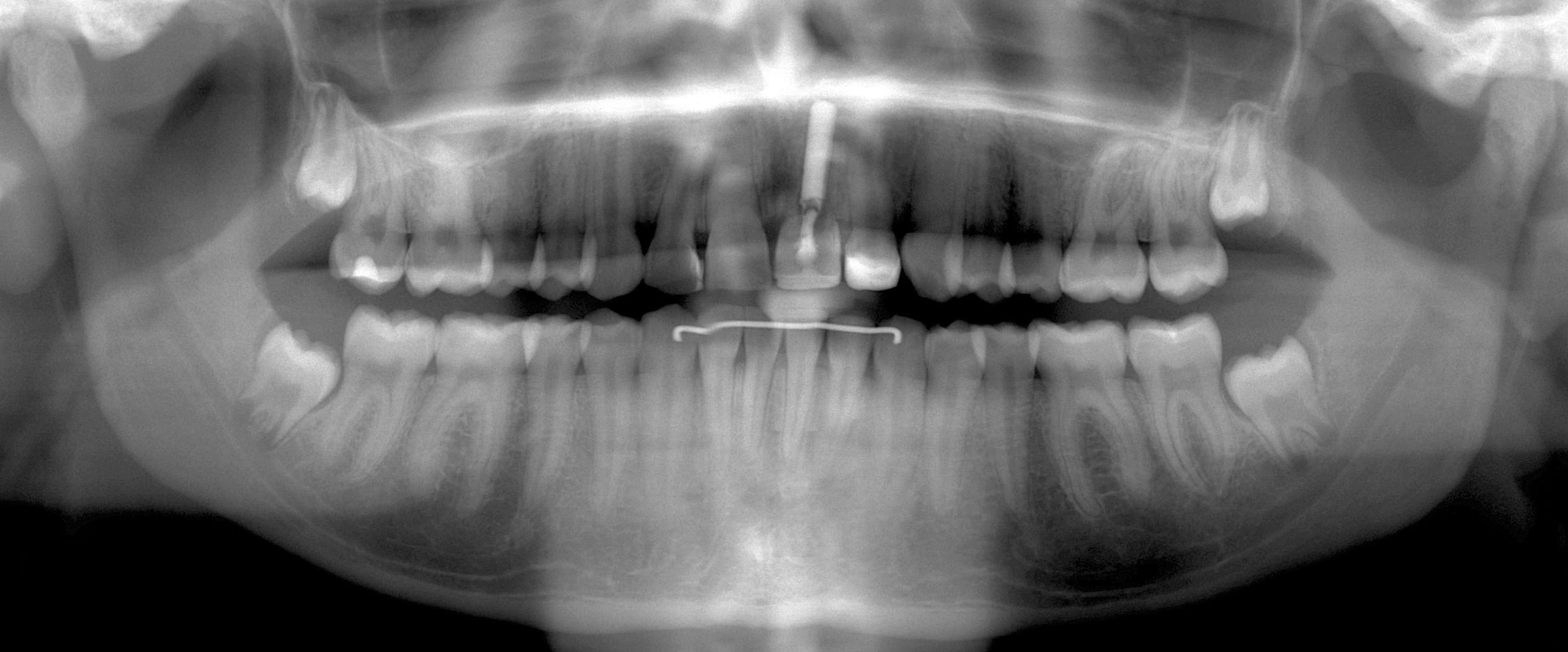
Figure 3
CBCT analysis of the site after extraction of the left maxillary central incisor including the titanium post (small FOV: 4 x 4 cm): the evaluation of the images exhibits a root canal filled left lateral incisor, a prominent nasopalatine canal (Y-shaped), and visible excessive root canal filling material in the bone apically to both maxillary left incisors.
Based on the findings of the CBCT images, a dental implant placement with simultaneous guided bone regeneration was planned.
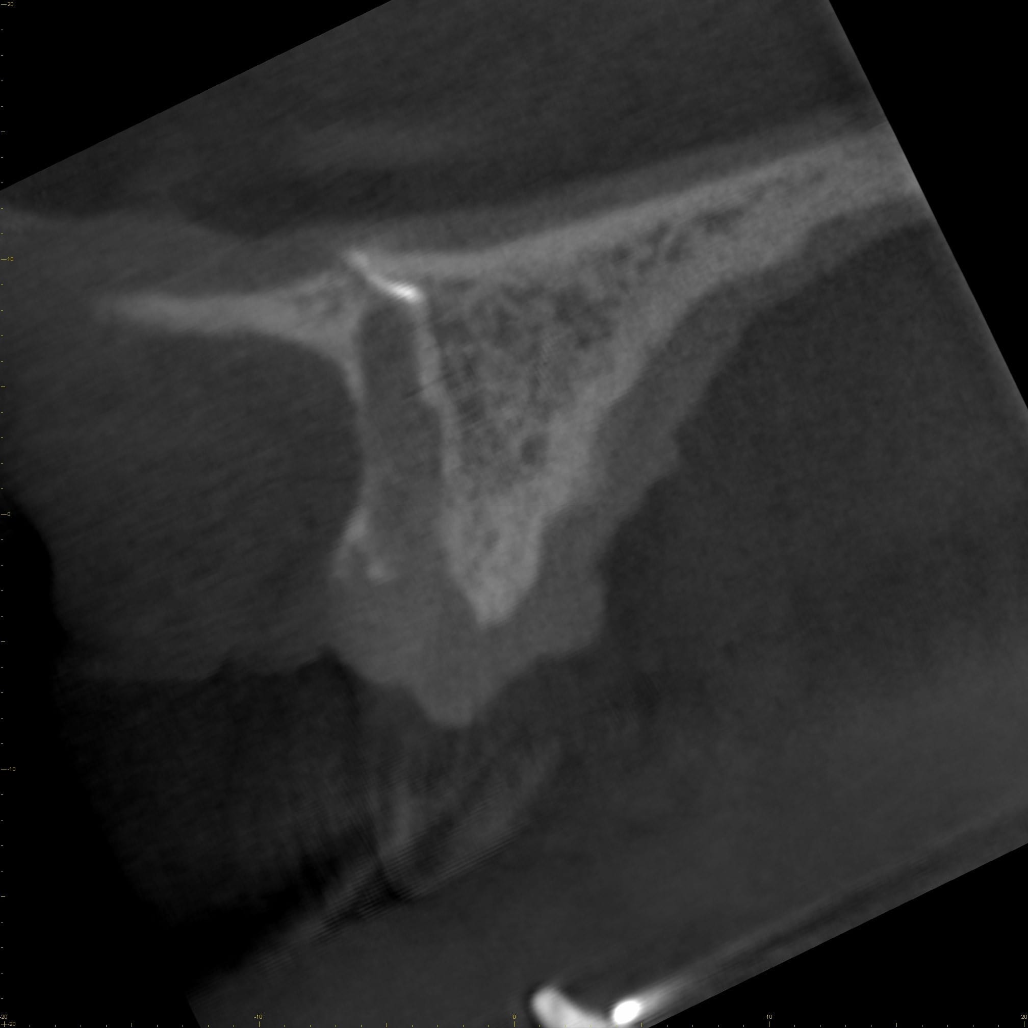
Figure 3A: Sagittal CBCT image
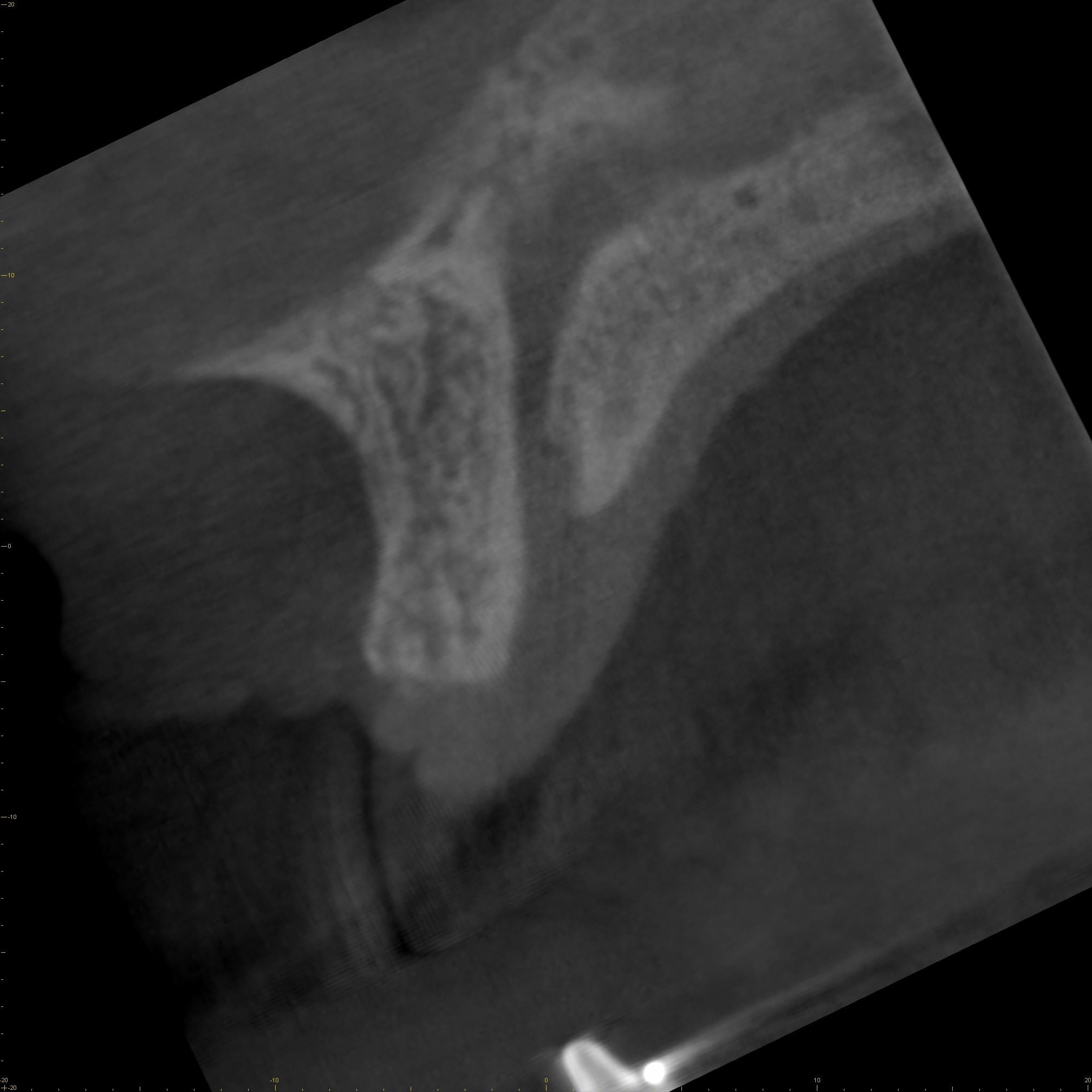
Figure 3B: Sagittal CBCT image
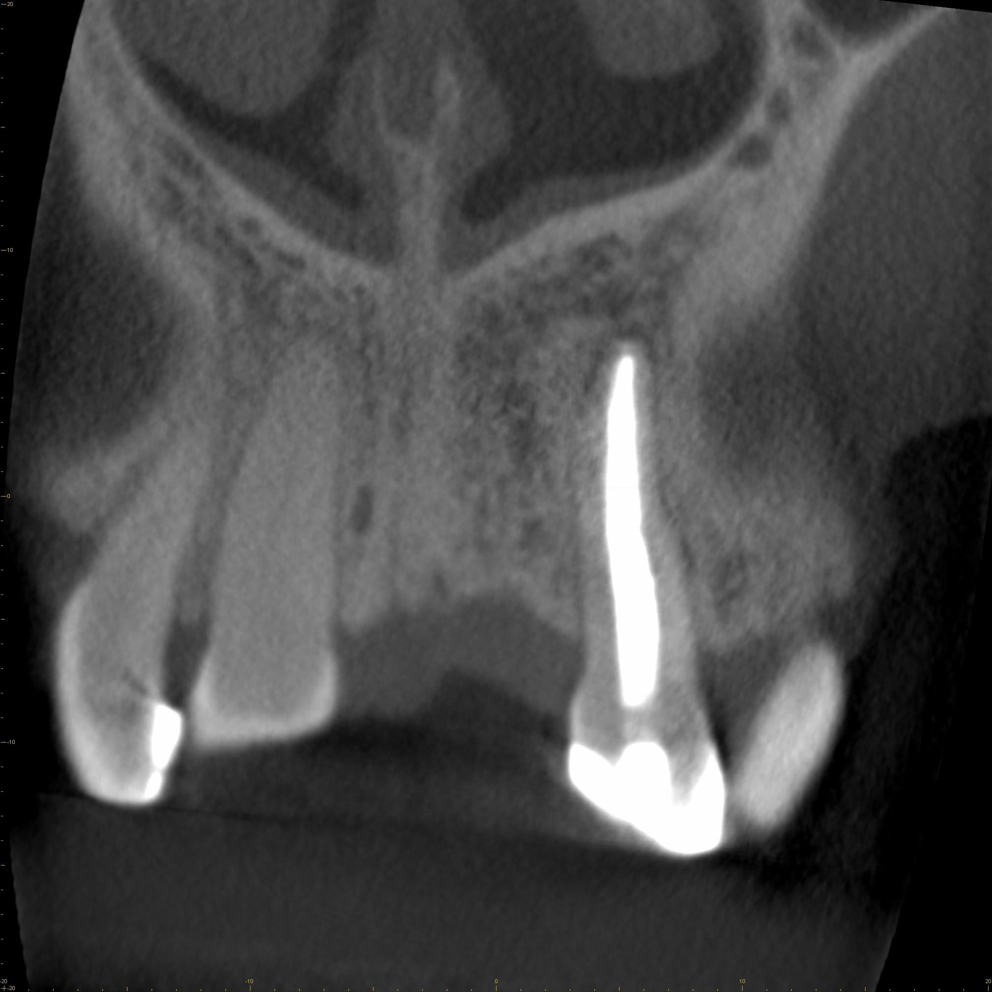
Figure 3C: Coronal CBCT image
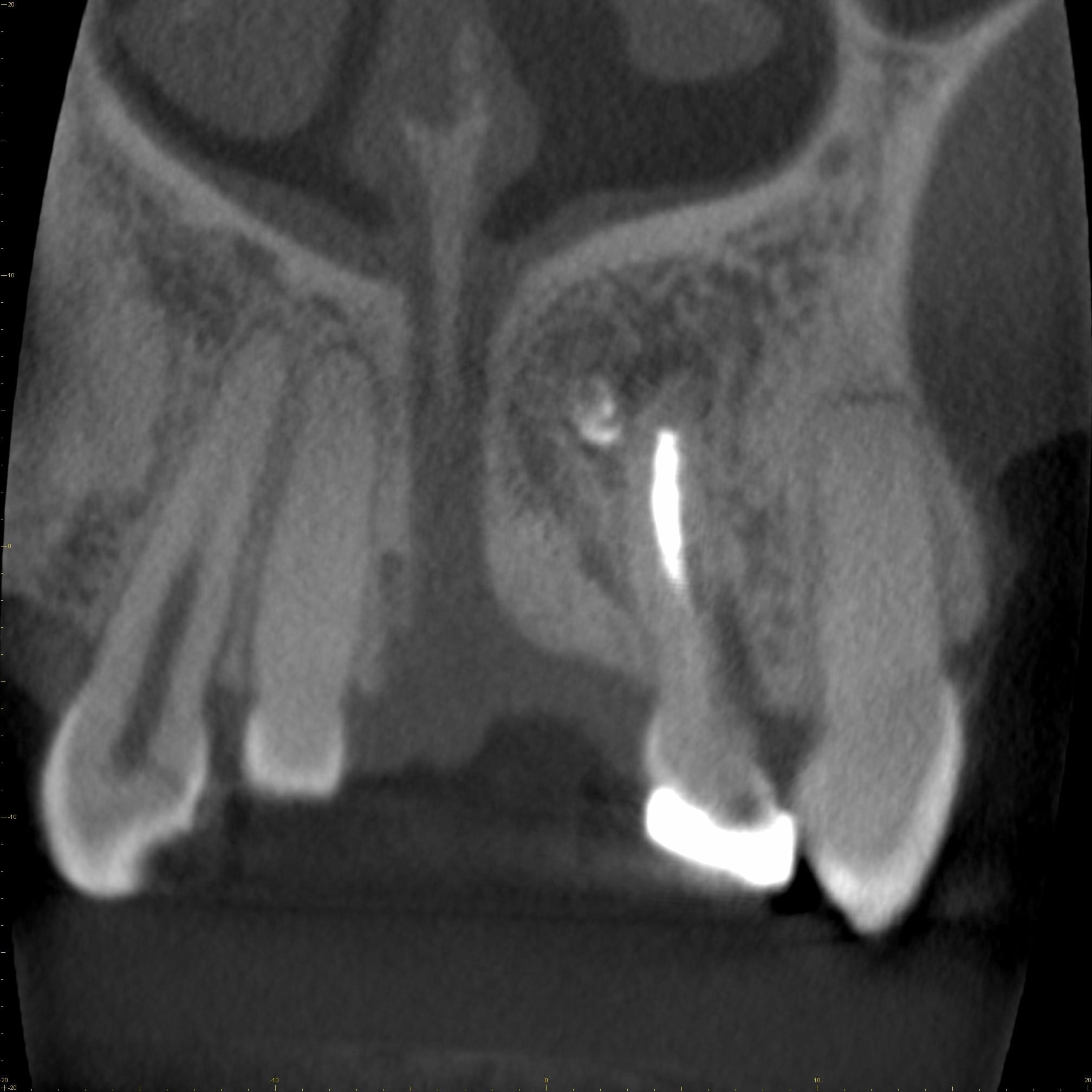 Figure 3D: Coronal CBCT image
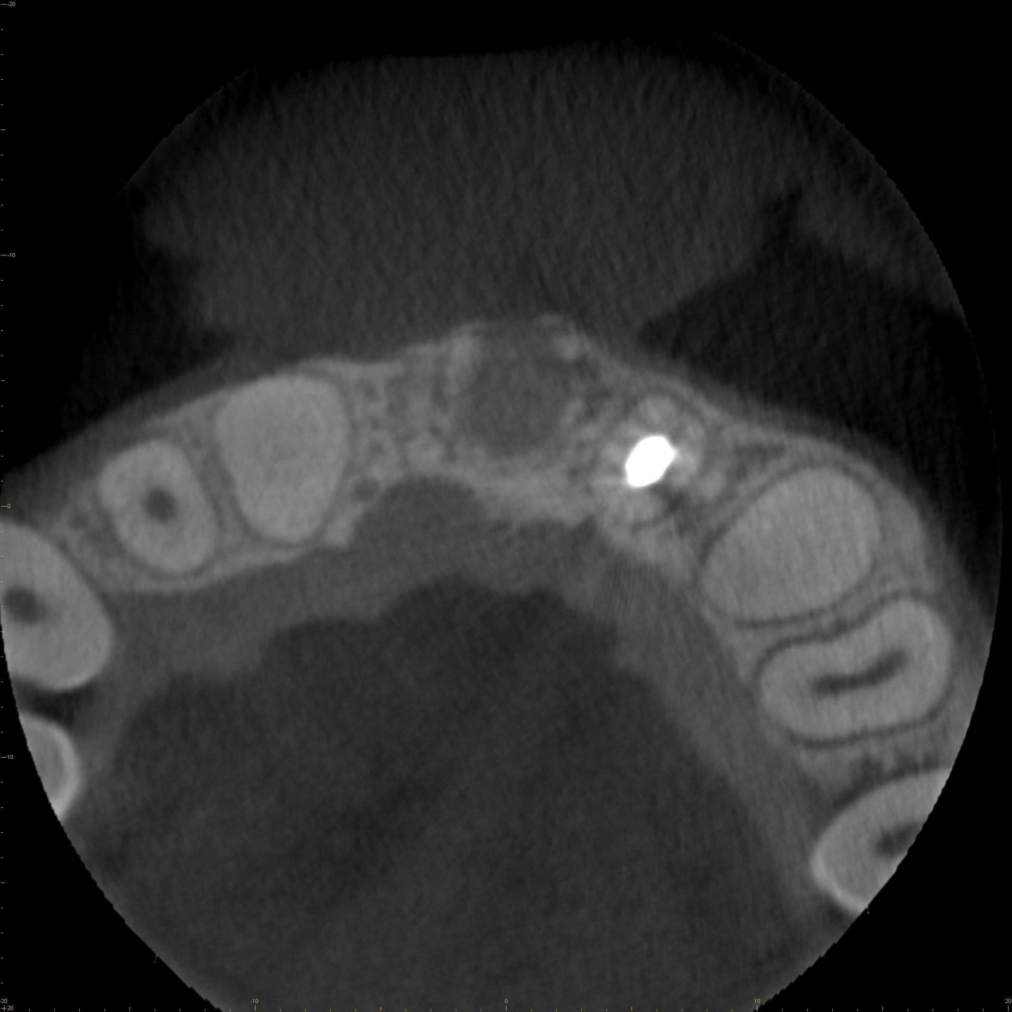 Figure 3E: Axial CBCT image
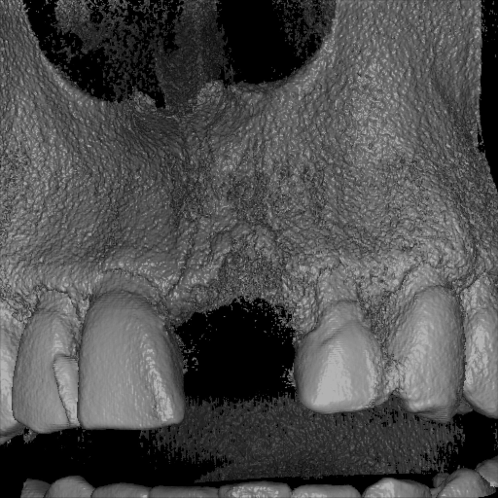 Figure 3F: Volume rendered data set
Figure 4
Radiographic (A) and clinical (B) presentation two years after dental implant insertion in the region of the left maxillary central incisor.
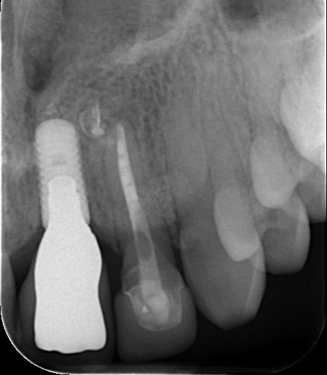
Figure 4A
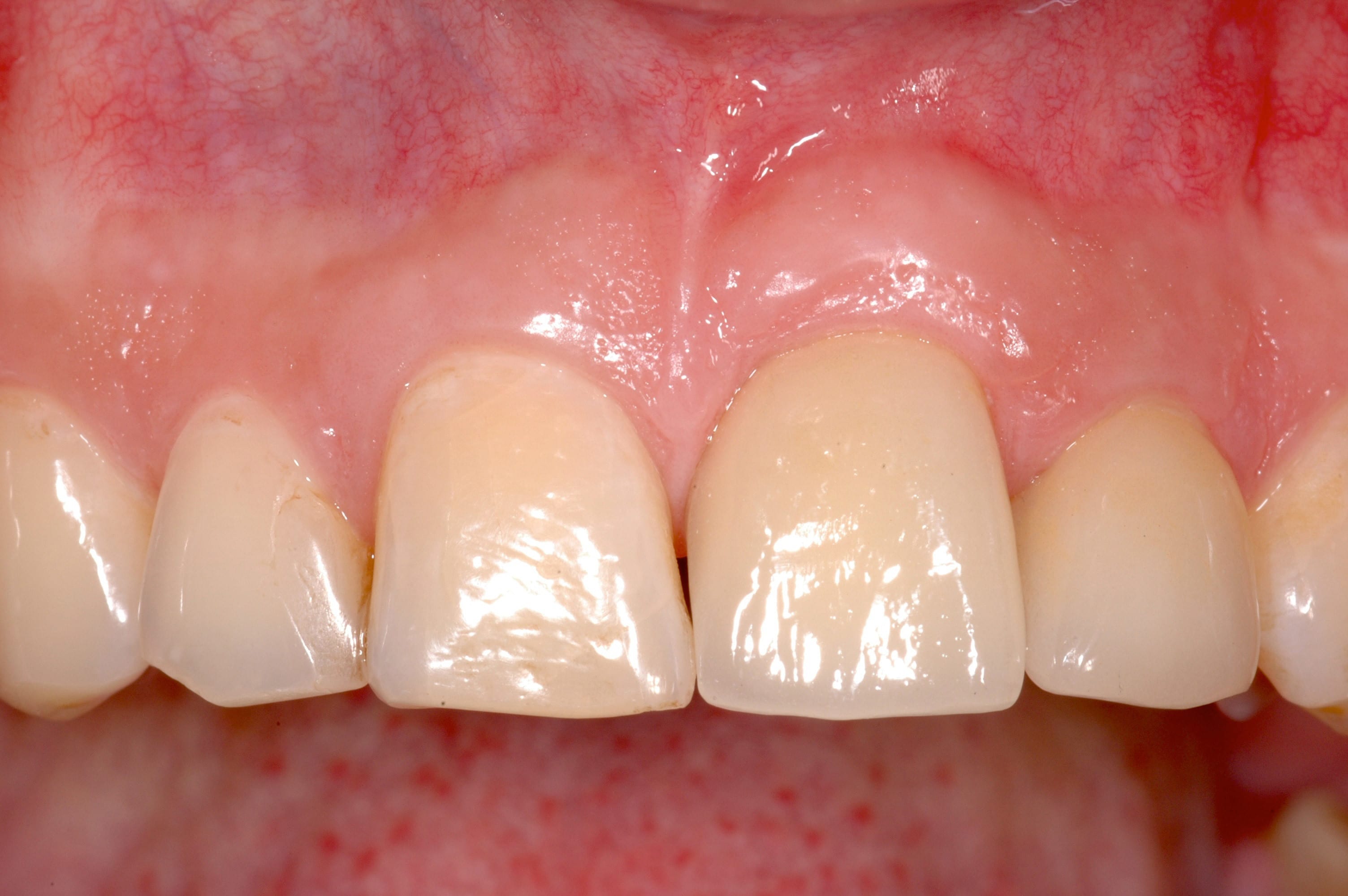
Figure 4B


