Figure 1
Panoramic view of a 64-year old patient
Panoramic view of a 64-year old patient exhibiting generalized bone loss due to periodontal disease including vertical defects in the left maxillary first molar and mandibular incisor area. The patient complained about mobile teeth in the anterior mandible and halitosis.
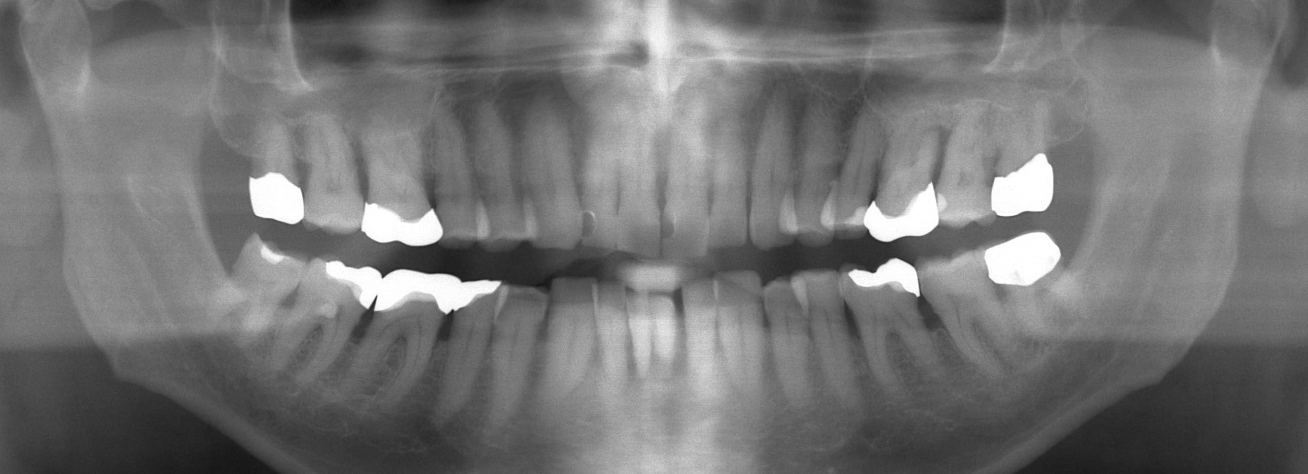
Figure 2
CBCT analysis and implant treatment planning in the anterior mandibular region following periodontal therapy including extraction of the left maxillary first molar and three mandibular incisors
The 3D images (small FOV: 6 x 6 cm) exhibit sufficient vertical and horizontal bone height for the placement of two dental implants with reduced diameter.
 Figure 2A: Coronal CBCT image with both canines laterally to the planned implants
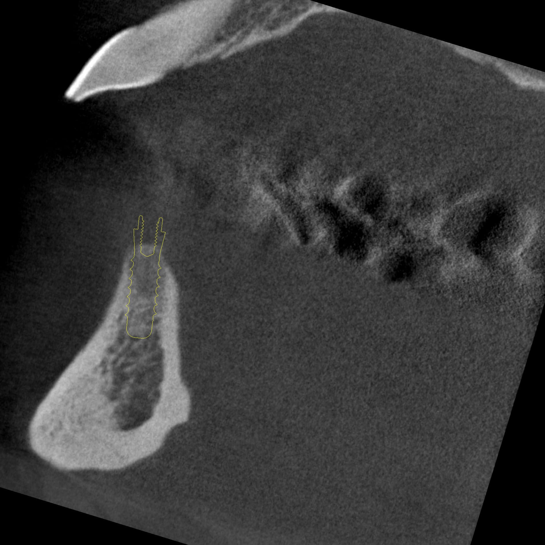 Figure 2B: Sagittal CBCT image for implants in the position of the right and left lateral incisor
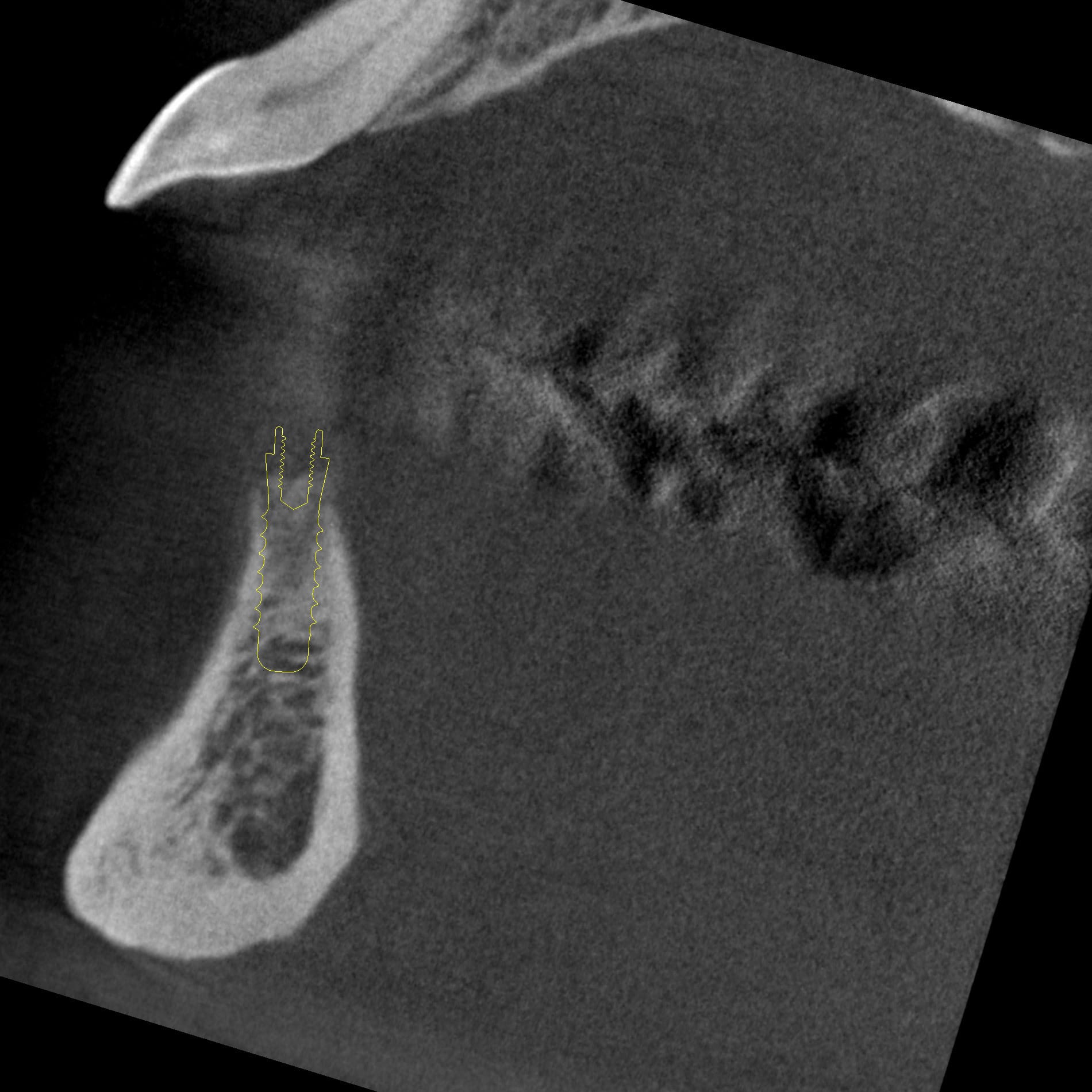 Figure 2C: Sagittal CBCT image for implants in the position of the right lateral incisor
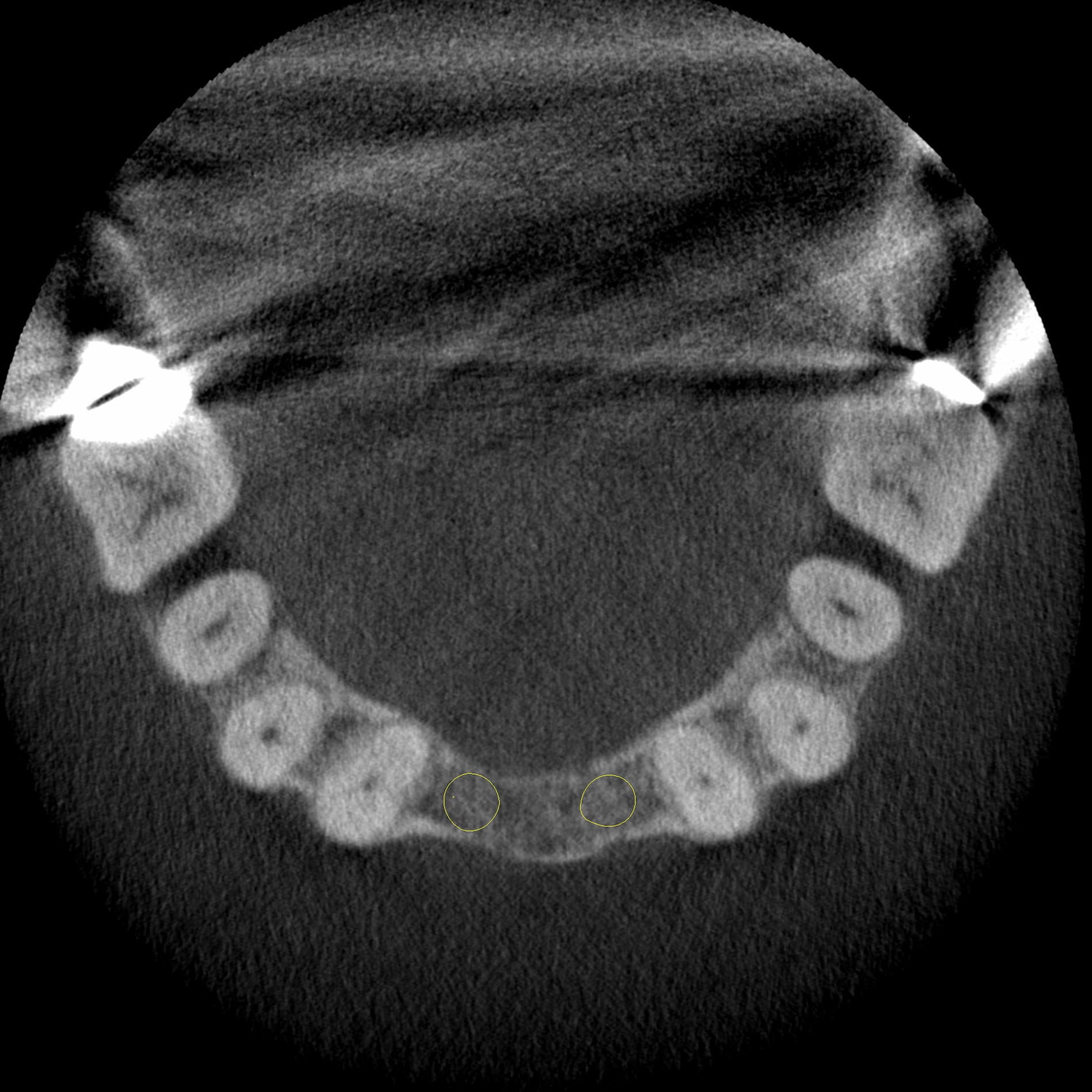 Figure 2D: Axial CBCT image at a coronal and apical position
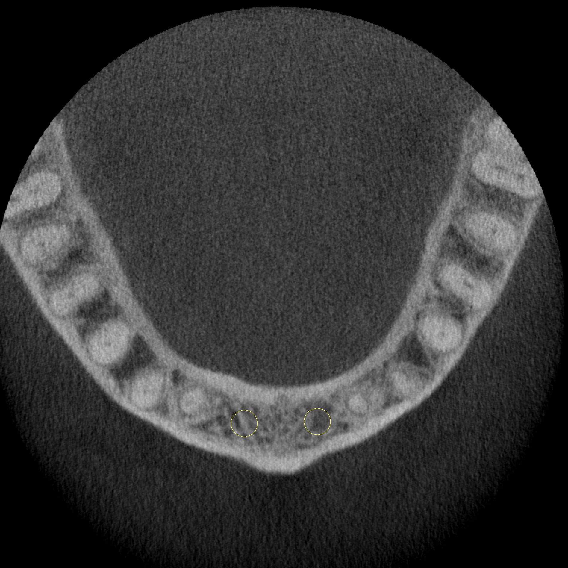 Figure 2E: Axial CBCT image at a coronal position
 Figure 2F: Volume rendered data set
Figure 3
Presentation two years after dental implant insertion in the region of the anterior mandible
The missing teeth were restored with a fixed partial denture of three units including pink porcelain to cover the bone and soft tissue loss due to initial periodontal breakdown.
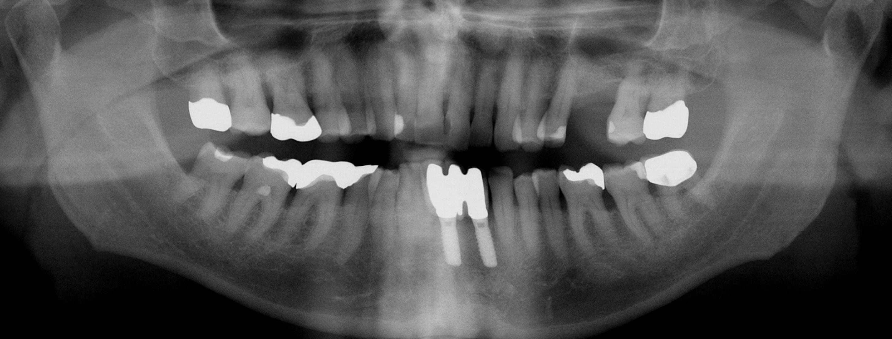 Figure 3A: Panoramic view
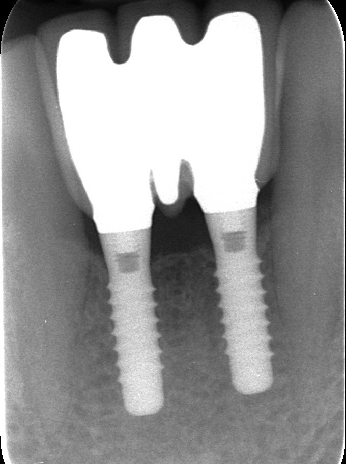 Figure 3B: Periapical radiograph
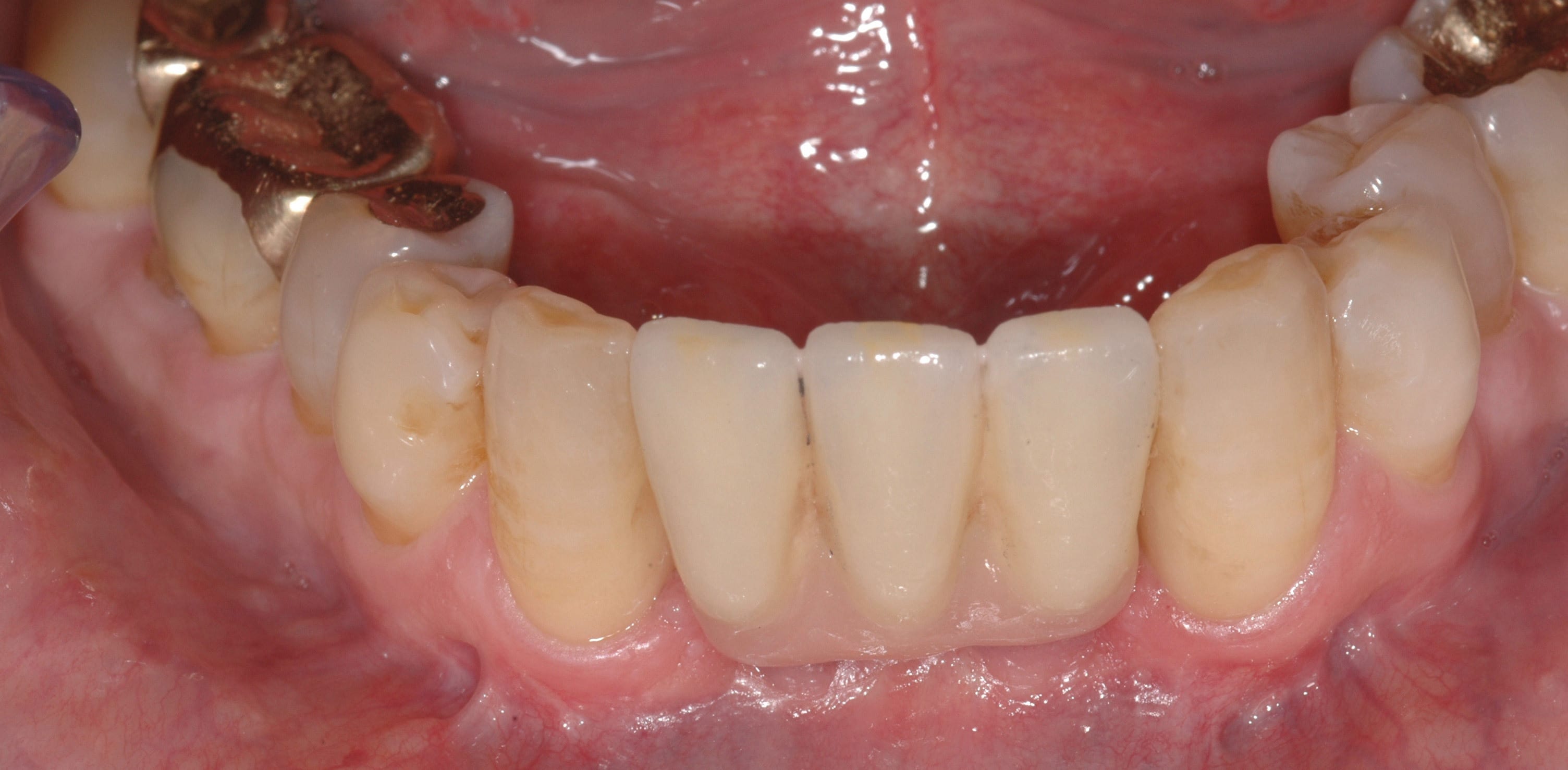 Figure 3C: Clinical aspect


